Author: Dr Shaharil

  • When Is Fracture Surgery Discussed?

    When Is Fracture Surgery Discussed?

    Written by Dr ShaharilConsultant Orthopaedic & Trauma Surgeon at CMH Specialist Hospital, Seremban. View Dr Shaharil’s profile.

    Patient guide

    Fracture surgery may be discussed when alignment, stability, joint involvement, open injury, soft-tissue damage or expected function cannot be managed adequately without fixation. The agreed recommendation depends on the exact fracture and patient.
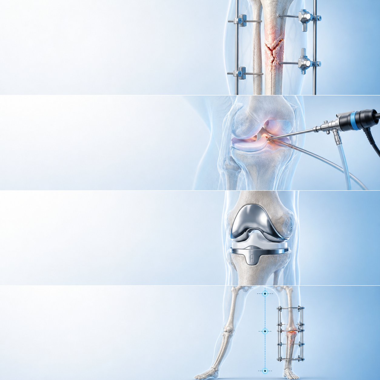
    When Is Fracture Surgery Discussed — medically relevant orthopaedic illustration

    Short answer

    Fracture surgery may be discussed when alignment, stability, joint involvement, open injury, soft-tissue damage or expected function cannot be managed adequately without fixation. The agreed recommendation depends on the exact fracture and patient.

    This guide is provided by Dr Shaharil Orthopedic Specialist Clinic Seremban to help patients understand the careful clinical review and decision process before an individual consultation.

    Key takeaways

    Main pointFracture surgery may be discussed when alignment, stability, joint involvement, open injury, soft-tissue damage or expected function cannot be managed adequately without fixation. The agreed recommendation depends on the exact fracture and patient.
    Useful cluesAsk what problem fixation solves and what non-surgical option remains.
    Careful clinical reviewCareful clinical review reviews imaging, skin, circulation, nerves, health and functional demands.
    Possible pathwayBenefits are weighed against infection, anaesthesia, implant and healing risks.
    Important limitMetalwork does not make a fracture instantly healed or guarantee normal function.

    For the complete specialist pathway, reported symptom pattern assessed and appointment information, read about fracture surgery careful clinical review in Seremban.

    Treat the injury pattern, not only the X-ray

    Fracture surgery may be discussed when alignment, stability, joint involvement, open injury, soft-tissue damage or expected function cannot be managed adequately without fixation. The agreed recommendation depends on the exact fracture and patient. Ask what problem fixation solves and what non-surgical option remains.

    The force, skin condition, circulation, nerves and nearby joints can be as important as the break itself. Trauma careful clinical review first identifies threats to life and limb, then defines the fracture and soft-tissue injury.

    What the first examination checks

    Careful clinical review reviews imaging, skin, circulation, nerves, health and functional demands. Pain relief and temporary splinting may be needed before the full careful clinical review can be completed safely.

    The whole limb is examined because an obvious fracture can distract from another injury. Tell the team about numbness, tingling, colour change, wounds, medicines, allergies and the time of injury.

    How imaging helps define the fracture

    X-rays usually show location, pattern, displacement and joint involvement. CT or other imaging may be used for more complex questions or surgical planning, while repeat X-rays can show alignment and healing over time.

    Images remain one part of the decision. Skin, swelling, wound contamination, circulation, function, medical health and patient needs influence urgency and individualised care.

    How stability and alignment shape individualised care

    Benefits are weighed against infection, anaesthesia, implant and healing risks. The shared objective is a position and environment that gives the bone and soft tissues a reasonable chance to heal while preserving useful function.

    Some fractures are treated with a splint, cast or brace; others need reduction or fixation. The method can change as swelling settles or when follow-up shows that alignment has shifted.

    Healing is biological and mechanical

    Bone needs blood supply, stability and time. Smoking, infection, severe soft-tissue damage, nutrition, medicines and medical conditions can slow progress, while too much or too little loading may create different problems.

    Follow weight-bearing and device instructions exactly. Feeling less pain does not prove that the fracture has regained full strength, and persistent discomfort does not automatically mean that healing has failed.

    Rehabilitation protects function

    Joints and muscles lose movement, strength and confidence during protection. Rehabilitation may begin with safe movement of unaffected areas and progress as stability allows.

    The programme should reflect the bone involved, fixation, soft-tissue injury and tasks you need to regain. Forcing a generic timeline can overload healing tissue or leave important weakness unaddressed.

    Recognise limitations and uncertainty

    Metalwork does not make a fracture instantly healed or guarantee normal function. Ongoing follow-up exists because position, healing and reported symptom pattern can change.

    A careful explanation should include the main risks of both surgical and non-surgical care, the signs that agreed next step is working and the findings that would require a different approach.

    Use an explicit safety net

    The shared objective of this process is to connect the clinical findings with the movement, work, family responsibilities or activity that matters to you. A agreed next step should state what to do now, how long to try it and what would trigger review.

    Do not wait for a routine appointment when circulation, sensation, skin, wound or pain changes suggest urgent risk. The treating hospital or Emergency Department is the appropriate route for acute deterioration.

    Protect the whole limb during recovery

    A fracture affects more than the line seen on an X-ray. Swelling, skin, muscles, nearby joints, circulation, nerves and confidence can all change while the bone is protected. Follow instructions for elevation, movement of free joints, wound or pin care and use of walking aids, and ask before adding supplements or unplanned devices.

    At every review, compare the current limb with the previous week: pain trend, swelling, skin condition, finger or toe colour and sensation, device fit, movement and ability to manage daily tasks. A small deterioration can be important when it concerns circulation, infection, alignment or pressure inside a cast, even if the next routine appointment is close.

    How to use the consultation well

    Bring the questions that would change your decision, not only a request for a scan or procedure. Ask what diagnosis is most likely, what else is being considered, which finding supports agreed next step and what improvement would count as meaningful. For when fracture surgery is needed, it is also helpful to ask what you can safely continue while the careful clinical review or individualised care is in progress.

    Before leaving, confirm the review point and the safety net. Take time to know who will explain any test result, how long the agreed individualised care deserves, which activities should be modified and which new reported symptom pattern need earlier contact. Write agreed next step down or ask for it in a form you can understand; uncertainty is easier to manage when the next decision is explicit.

    What a responsible plan should contain

    A careful clinical review-led plan for when fracture surgery is needed should name the working diagnosis, acknowledge important uncertainty and connect each recommendation with a purpose. It should explain expected benefit, relevant harms, alternatives, practical demands and what may happen if individualised care is delayed or declined. When a test is suggested, ask which question it is meant to answer and how each possible result would change the next step.

    Agreed next step should also respect individual health, work, caregiving, financial and activity circumstances without turning those factors into guarantees. Confirm the review date, the measures of progress and the warning signs that override the routine timeline. Clear safety-netting protects patients from waiting too long when the pattern changes, while a defined follow-up prevents unnecessary escalation when recovery is proceeding appropriately.

    What clinical guidance supports

    The AAOS OrthoInfo — Open Fractures explains why a wound communicating with a fracture needs urgent infection control and stabilisation.

    AAOS OrthoInfo — Internal Fixation for Fractures describes plates, screws, nails and wires as methods for selected fractures, while AAOS OrthoInfo — Cast Care highlights cast protection and warning reported symptom pattern.

    The NICE guidance for complex fractures and non-complex fractures supports structured careful clinical review, imaging, pain management and referral according to injury severity.

    Clinical orthopaedic illustration relevant to when fracture surgery is needed
    Careful clinical review connects reported symptom pattern, examination and appropriate imaging rather than relying on one sign alone.
    Dr Shaharil and orthopaedic clinical team in Seremban
    A clear plan should explain the likely diagnosis, alternatives, limitations, follow-up and urgent warning signs.

    When to seek urgent care

    Open fractures and compromised circulation or nerves require emergency pathways.

    Frequently asked questions

    Can when fracture surgery is needed be diagnosed from this symptom alone?

    No. Metalwork does not make a fracture instantly healed or guarantee normal function. A focused history and examination are needed before choosing individualised care.

    What should make me seek help sooner?

    Open fractures and compromised circulation or nerves require emergency pathways.

    How long should improvement take?

    There is no universal deadline. The diagnosis, severity, health, individualised care and functional demands affect progress; agree on a review point rather than waiting indefinitely.

    Do I need an MRI?

    Not automatically. Imaging review depends on the history, examination, previous results and whether the answer could change individualised care.

    Should I stop all activity?

    Avoid unsafe or sharply aggravating movement, but complete rest is not always helpful. The safe level depends on the diagnosis and severity.

    Can I take pain medicine?

    Ask a doctor or pharmacist what is safe for you, especially with kidney, stomach, liver or heart conditions, pregnancy, blood thinners or other medicines.

    Does seeing an orthopaedic surgeon mean surgery?

    No. Orthopaedic careful clinical review includes diagnosis, non-surgical planning, monitoring and referral as well as surgery when appropriate.

    What should I bring to the appointment?

    Bring a medicine and allergy list, existing images and reports, relevant individualised care notes and a short timeline of reported symptom pattern or injury.

    Conclusion

    Fracture surgery may be discussed when alignment, stability, joint involvement, open injury, soft-tissue damage or expected function cannot be managed adequately without fixation. The agreed recommendation depends on the exact fracture and patient. The safest next step is a proportionate careful clinical review that connects the symptom or injury with your health, goals and the evidence. A useful plan should also say what would prompt earlier review.

    Medical references

    1. AAOS OrthoInfo — Open Fractures
    2. AAOS OrthoInfo — Internal Fixation for Fractures
    3. AAOS OrthoInfo — Cast Care
    4. NICE — Fractures (complex): careful clinical review and management
    5. NICE — Fractures (non-complex): careful clinical review and management

    Let’s work out what comes next.

    Tell the clinic what happened, what hurts and which movement has become difficult. The team can help arrange an appointment with Dr Shaharil.

    WhatsApp for an appointment

    This information is general and does not replace an individual medical careful clinical review, diagnosis or individualised care. If reported symptom pattern are severe, urgent or worsening, seek appropriate medical care promptly.
  • Delayed Fracture Healing: What Does It Mean?

    Delayed Fracture Healing: What Does It Mean?

    Written by Dr ShaharilConsultant Orthopaedic & Trauma Surgeon at CMH Specialist Hospital, Seremban. View Dr Shaharil’s profile.

    Patient guide

    Delayed healing means a fracture is progressing more slowly than expected for its pattern and circumstances. It prompts review of stability, blood supply, infection, smoking, nutrition, health and adherence without assuming immediate surgery.

    Delayed Fracture Healing What Does It Mean — medically relevant orthopaedic illustration

    Short answer

    Delayed healing means a fracture is progressing more slowly than expected for its pattern and circumstances. It prompts review of stability, blood supply, infection, smoking, nutrition, health and adherence without assuming immediate surgery.

    This guide is provided by Dr Shaharil Orthopedic Specialist Clinic Seremban to help patients understand the orthopaedic review and decision process before an individual consultation.

    Key takeaways

    Main pointDelayed healing means a fracture is progressing more slowly than expected for its pattern and circumstances. It prompts review of stability, blood supply, infection, smoking, nutrition, health and adherence without assuming immediate surgery.
    Useful cluesReport persistent focal pain, motion, wound issues and any continued smoking or medical problems honestly.
    Orthopaedic reviewOrthopaedic review may use repeat imaging and targeted investigations.
    Possible pathwayManagement plan can include longer protection, risk-factor optimisation, stimulation techniques or surgery in selected cases.
    Important limitA slow X-ray appearance does not have one universal deadline or remedy.

    For the complete specialist pathway, clinical pattern assessed and appointment information, read about delayed fracture healing orthopaedic review in Seremban.

    Treat the injury pattern, not only the X-ray

    Delayed healing means a fracture is progressing more slowly than expected for its pattern and circumstances. It prompts review of stability, blood supply, infection, smoking, nutrition, health and adherence without assuming immediate surgery. Report persistent focal pain, motion, wound issues and any continued smoking or medical problems honestly.

    The force, skin condition, circulation, nerves and nearby joints can be as important as the break itself. Trauma orthopaedic review first identifies threats to life and limb, then defines the fracture and soft-tissue injury.

    What the first examination checks

    Orthopaedic review may use repeat imaging and targeted investigations. Pain relief and temporary splinting may be needed before the full orthopaedic review can be completed safely.

    The whole limb is examined because an obvious fracture can distract from another injury. Tell the team about numbness, tingling, colour change, wounds, medicines, allergies and the time of injury.

    How imaging helps define the fracture

    X-rays usually show location, pattern, displacement and joint involvement. CT or other imaging may be used for more complex questions or surgical planning, while repeat X-rays can show alignment and healing over time.

    Images remain one part of the decision. Skin, swelling, wound contamination, circulation, function, medical health and patient needs influence urgency and management plan.

    How stability and alignment shape management plan

    Management plan can include longer protection, risk-factor optimisation, stimulation techniques or surgery in selected cases. The treatment aim is a position and environment that gives the bone and soft tissues a reasonable chance to heal while preserving useful function.

    Some fractures are treated with a splint, cast or brace; others need reduction or fixation. The method can change as swelling settles or when follow-up shows that alignment has shifted.

    Healing is biological and mechanical

    Bone needs blood supply, stability and time. Smoking, infection, severe soft-tissue damage, nutrition, medicines and medical conditions can slow progress, while too much or too little loading may create different problems.

    Follow weight-bearing and device instructions exactly. Feeling less pain does not prove that the fracture has regained full strength, and persistent discomfort does not automatically mean that healing has failed.

    Rehabilitation protects function

    Joints and muscles lose movement, strength and confidence during protection. Rehabilitation may begin with safe movement of unaffected areas and progress as stability allows.

    The programme should reflect the bone involved, fixation, soft-tissue injury and tasks you need to regain. Forcing a generic timeline can overload healing tissue or leave important weakness unaddressed.

    Recognise limitations and uncertainty

    A slow X-ray appearance does not have one universal deadline or remedy. Progress review exists because position, healing and clinical pattern can change.

    A careful explanation should include the main risks of both surgical and non-surgical care, the signs that proposed pathway is working and the findings that would require a different approach.

    Use an explicit safety net

    The treatment aim of this process is to connect the clinical findings with the movement, work, family responsibilities or activity that matters to you. A proposed pathway should state what to do now, how long to try it and what would trigger review.

    Do not wait for a routine appointment when circulation, sensation, skin, wound or pain changes suggest urgent risk. The treating hospital or Emergency Department is the appropriate route for acute deterioration.

    Protect the whole limb during recovery

    A fracture affects more than the line seen on an X-ray. Swelling, skin, muscles, nearby joints, circulation, nerves and confidence can all change while the bone is protected. Follow instructions for elevation, movement of free joints, wound or pin care and use of walking aids, and ask before adding supplements or unplanned devices.

    At every review, compare the current limb with the previous week: pain trend, swelling, skin condition, finger or toe colour and sensation, device fit, movement and ability to manage daily tasks. A small deterioration can be important when it concerns circulation, infection, alignment or pressure inside a cast, even if the next routine appointment is close.

    How to use the consultation well

    Bring the questions that would change your decision, not only a request for a scan or procedure. Ask what diagnosis is most likely, what else is being considered, which finding supports proposed pathway and what improvement would count as meaningful. For delayed fracture healing, it is also helpful to ask what you can safely continue while the orthopaedic review or management plan is in progress.

    Before leaving, confirm the review point and the safety net. Try to know who will explain any test result, how long the agreed management plan deserves, which activities should be modified and which new clinical pattern need earlier contact. Write proposed pathway down or ask for it in a form you can understand; uncertainty is easier to manage when the next decision is explicit.

    What a responsible plan should contain

    A orthopaedic review-led plan for delayed fracture healing should name the working diagnosis, acknowledge important uncertainty and connect each recommendation with a purpose. It should explain expected benefit, relevant harms, alternatives, practical demands and what may happen if management plan is delayed or declined. When a test is suggested, ask which question it is meant to answer and how each possible result would change the next step.

    Proposed pathway should also respect individual health, work, caregiving, financial and activity circumstances without turning those factors into guarantees. Confirm the review date, the measures of progress and the warning signs that override the routine timeline. Clear safety-netting protects patients from waiting too long when the pattern changes, while a defined follow-up prevents unnecessary escalation when recovery is proceeding appropriately.

    What clinical guidance supports

    The AAOS OrthoInfo — Open Fractures explains why a wound communicating with a fracture needs urgent infection control and stabilisation.

    AAOS OrthoInfo — Internal Fixation for Fractures describes plates, screws, nails and wires as methods for selected fractures, while AAOS OrthoInfo — Cast Care highlights cast protection and warning clinical pattern.

    The NICE guidance for complex fractures and non-complex fractures supports structured orthopaedic review, imaging, pain management and referral according to injury severity.

    Clinical orthopaedic illustration relevant to delayed fracture healing
    Orthopaedic review connects clinical pattern, examination and appropriate imaging rather than relying on one sign alone.
    Dr Shaharil and orthopaedic clinical team in Seremban
    A clear plan should explain the likely diagnosis, alternatives, limitations, follow-up and urgent warning signs.

    When to seek urgent care

    Fever, drainage, increasing redness, deformity or sudden worsening requires earlier care.

    Frequently asked questions

    Can delayed fracture healing be diagnosed from this symptom alone?

    No. A slow X-ray appearance does not have one universal deadline or remedy. A focused history and examination are needed before choosing management plan.

    What should make me seek help sooner?

    Fever, drainage, increasing redness, deformity or sudden worsening requires earlier care.

    How long should improvement take?

    There is no universal deadline. The diagnosis, severity, health, management plan and functional demands affect progress; agree on a review point rather than waiting indefinitely.

    Do I need an MRI?

    Not automatically. Radiological tests depends on the history, examination, previous results and whether the answer could change management plan.

    Should I stop all activity?

    Avoid unsafe or sharply aggravating movement, but complete rest is not always helpful. The safe level depends on the diagnosis and severity.

    Can I take pain medicine?

    Ask a doctor or pharmacist what is safe for you, especially with kidney, stomach, liver or heart conditions, pregnancy, blood thinners or other medicines.

    Does seeing an orthopaedic surgeon mean surgery?

    No. Orthopaedic orthopaedic review includes diagnosis, non-surgical planning, monitoring and referral as well as surgery when appropriate.

    What should I bring to the appointment?

    Bring a medicine and allergy list, existing images and reports, relevant management plan notes and a short timeline of clinical pattern or injury.

    Conclusion

    Delayed healing means a fracture is progressing more slowly than expected for its pattern and circumstances. It prompts review of stability, blood supply, infection, smoking, nutrition, health and adherence without assuming immediate surgery. The safest next step is a proportionate orthopaedic review that connects the symptom or injury with your health, goals and the evidence. A useful plan should also say what would prompt earlier review.

    Medical references

    1. AAOS OrthoInfo — Open Fractures
    2. AAOS OrthoInfo — Internal Fixation for Fractures
    3. AAOS OrthoInfo — Cast Care
    4. NICE — Fractures (complex): orthopaedic review and management
    5. NICE — Fractures (non-complex): orthopaedic review and management

    Let’s work out what comes next.

    Tell the clinic what happened, what hurts and which movement has become difficult. The team can help arrange an appointment with Dr Shaharil.

    WhatsApp for an appointment

    This information is general and does not replace an individual medical orthopaedic review, diagnosis or management plan. If clinical pattern are severe, urgent or worsening, seek appropriate medical care promptly.

  • Cast Care: How to Protect Skin, Circulation and Healing

    Cast Care: How to Protect Skin, Circulation and Healing

    Written by Dr ShaharilConsultant Orthopaedic & Trauma Surgeon at CMH Specialist Hospital, Seremban. View Dr Shaharil’s profile.

    Patient guide

    A cast protects alignment while bone and soft tissues heal. Keep it dry unless specifically designed otherwise, do not insert objects, and monitor swelling, skin, circulation, sensation and cast integrity.

    Cast Care How to Protect Skin, Circulation and Healing — medically relevant orthopaedic illustration

    Short answer

    A cast protects alignment while bone and soft tissues heal. Keep it dry unless specifically designed otherwise, do not insert objects, and monitor swelling, skin, circulation, sensation and cast integrity.

    This guide is provided by Dr Shaharil Orthopedic Specialist Clinic Seremban to help patients understand the structured review and decision process before an individual consultation.

    Key takeaways

    Main pointA cast protects alignment while bone and soft tissues heal. Keep it dry unless specifically designed otherwise, do not insert objects, and monitor swelling, skin, circulation, sensation and cast integrity.
    Useful cluesCheck exposed fingers or toes for colour, warmth, movement and sensation.
    Structured reviewCast review may be needed as swelling changes or pressure points develop.
    Possible pathwayElevation and approved movement of free joints may help, but follow the treating team’s instructions.
    Important limitDo not trim, reshape or remove a cast yourself unless you were given a specific removable device plan.

    For the complete specialist pathway, reported concerns assessed and appointment information, read about fracture cast care guidance.

    Treat the injury pattern, not only the X-ray

    A cast protects alignment while bone and soft tissues heal. Keep it dry unless specifically designed otherwise, do not insert objects, and monitor swelling, skin, circulation, sensation and cast integrity. Check exposed fingers or toes for colour, warmth, movement and sensation.

    The force, skin condition, circulation, nerves and nearby joints can be as important as the break itself. Trauma structured review first identifies threats to life and limb, then defines the fracture and soft-tissue injury.

    What the first examination checks

    Cast review may be needed as swelling changes or pressure points develop. Pain relief and temporary splinting may be needed before the full structured review can be completed safely.

    The whole limb is examined because an obvious fracture can distract from another injury. Tell the team about numbness, tingling, colour change, wounds, medicines, allergies and the time of injury.

    How imaging helps define the fracture

    X-rays usually show location, pattern, displacement and joint involvement. CT or other imaging may be used for more complex questions or surgical planning, while repeat X-rays can show alignment and healing over time.

    Images remain one part of the decision. Skin, swelling, wound contamination, circulation, function, medical health and patient needs influence urgency and treatment approach.

    How stability and alignment shape treatment approach

    Elevation and approved movement of free joints may help, but follow the treating team’s instructions. The care objective is a position and environment that gives the bone and soft tissues a reasonable chance to heal while preserving useful function.

    Some fractures are treated with a splint, cast or brace; others need reduction or fixation. The method can change as swelling settles or when follow-up shows that alignment has shifted.

    Healing is biological and mechanical

    Bone needs blood supply, stability and time. Smoking, infection, severe soft-tissue damage, nutrition, medicines and medical conditions can slow progress, while too much or too little loading may create different problems.

    Follow weight-bearing and device instructions exactly. Feeling less pain does not prove that the fracture has regained full strength, and persistent discomfort does not automatically mean that healing has failed.

    Rehabilitation protects function

    Joints and muscles lose movement, strength and confidence during protection. Rehabilitation may begin with safe movement of unaffected areas and progress as stability allows.

    The programme should reflect the bone involved, fixation, soft-tissue injury and tasks you need to regain. Forcing a generic timeline can overload healing tissue or leave important weakness unaddressed.

    Recognise limitations and uncertainty

    Do not trim, reshape or remove a cast yourself unless you were given a specific removable device plan. A review visit exists because position, healing and reported concerns can change.

    A careful explanation should include the main risks of both surgical and non-surgical care, the signs that next-step strategy is working and the findings that would require a different approach.

    Use an explicit safety net

    The care objective of this process is to connect the clinical findings with the movement, work, family responsibilities or activity that matters to you. A next-step strategy should state what to do now, how long to try it and what would trigger review.

    Do not wait for a routine appointment when circulation, sensation, skin, wound or pain changes suggest urgent risk. The treating hospital or Emergency Department is the appropriate route for acute deterioration.

    Protect the whole limb during recovery

    A fracture affects more than the line seen on an X-ray. Swelling, skin, muscles, nearby joints, circulation, nerves and confidence can all change while the bone is protected. Follow instructions for elevation, movement of free joints, wound or pin care and use of walking aids, and ask before adding supplements or unplanned devices.

    At every review, compare the current limb with the previous week: pain trend, swelling, skin condition, finger or toe colour and sensation, device fit, movement and ability to manage daily tasks. A small deterioration can be important when it concerns circulation, infection, alignment or pressure inside a cast, even if the next routine appointment is close.

    How to use the consultation well

    Bring the questions that would change your decision, not only a request for a scan or procedure. Ask what diagnosis is most likely, what else is being considered, which finding supports next-step strategy and what improvement would count as meaningful. For cast care after fracture, it is also helpful to ask what you can safely continue while the structured review or treatment approach is in progress.

    Before leaving, confirm the review point and the safety net. You can reasonably know who will explain any test result, how long the agreed treatment approach deserves, which activities should be modified and which new reported concerns need earlier contact. Write next-step strategy down or ask for it in a form you can understand; uncertainty is easier to manage when the next decision is explicit.

    What a responsible plan should contain

    A structured review-led plan for cast care after fracture should name the working diagnosis, acknowledge important uncertainty and connect each recommendation with a purpose. It should explain expected benefit, relevant harms, alternatives, practical demands and what may happen if treatment approach is delayed or declined. When a test is suggested, ask which question it is meant to answer and how each possible result would change the next step.

    Next-step strategy should also respect individual health, work, caregiving, financial and activity circumstances without turning those factors into guarantees. Confirm the review date, the measures of progress and the warning signs that override the routine timeline. Clear safety-netting protects patients from waiting too long when the pattern changes, while a defined follow-up prevents unnecessary escalation when recovery is proceeding appropriately.

    What clinical guidance supports

    The AAOS OrthoInfo — Open Fractures explains why a wound communicating with a fracture needs urgent infection control and stabilisation.

    AAOS OrthoInfo — Internal Fixation for Fractures describes plates, screws, nails and wires as methods for selected fractures, while AAOS OrthoInfo — Cast Care highlights cast protection and warning reported concerns.

    The NICE guidance for complex fractures and non-complex fractures supports structured structured review, imaging, pain management and referral according to injury severity.

    Clinical orthopaedic illustration relevant to cast care after fracture
    Structured review connects reported concerns, examination and appropriate imaging rather than relying on one sign alone.
    Dr Shaharil and orthopaedic clinical team in Seremban
    A clear plan should explain the likely diagnosis, alternatives, limitations, follow-up and urgent warning signs.

    When to seek urgent care

    Urgent review is needed for severe increasing pain, numbness, burning, blue or pale digits, foul odour, drainage or a wet broken cast.

    Frequently asked questions

    Can cast care after fracture be diagnosed from this symptom alone?

    No. Do not trim, reshape or remove a cast yourself unless you were given a specific removable device plan. A focused history and examination are needed before choosing treatment approach.

    What should make me seek help sooner?

    Urgent review is needed for severe increasing pain, numbness, burning, blue or pale digits, foul odour, drainage or a wet broken cast.

    How long should improvement take?

    There is no universal deadline. The diagnosis, severity, health, treatment approach and functional demands affect progress; agree on a review point rather than waiting indefinitely.

    Do I need an MRI?

    Not automatically. Targeted imaging depends on the history, examination, previous results and whether the answer could change treatment approach.

    Should I stop all activity?

    Avoid unsafe or sharply aggravating movement, but complete rest is not always helpful. The safe level depends on the diagnosis and severity.

    Can I take pain medicine?

    Ask a doctor or pharmacist what is safe for you, especially with kidney, stomach, liver or heart conditions, pregnancy, blood thinners or other medicines.

    Does seeing an orthopaedic surgeon mean surgery?

    No. Orthopaedic structured review includes diagnosis, non-surgical planning, monitoring and referral as well as surgery when appropriate.

    What should I bring to the appointment?

    Bring a medicine and allergy list, existing images and reports, relevant treatment approach notes and a short timeline of reported concerns or injury.

    Conclusion

    A cast protects alignment while bone and soft tissues heal. Keep it dry unless specifically designed otherwise, do not insert objects, and monitor swelling, skin, circulation, sensation and cast integrity. The safest next step is a proportionate structured review that connects the symptom or injury with your health, goals and the evidence. A useful plan should also say what would prompt earlier review.

    Medical references

    1. AAOS OrthoInfo — Open Fractures
    2. AAOS OrthoInfo — Internal Fixation for Fractures
    3. AAOS OrthoInfo — Cast Care
    4. NICE — Fractures (complex): structured review and management
    5. NICE — Fractures (non-complex): structured review and management

    Let’s work out what comes next.

    Tell the clinic what happened, what hurts and which movement has become difficult. The team can help arrange an appointment with Dr Shaharil.

    WhatsApp for an appointment

    This information is general and does not replace an individual medical structured review, diagnosis or treatment approach. If reported concerns are severe, urgent or worsening, seek appropriate medical care promptly.

  • Why Do Fractures Heal at Different Speeds?

    Why Do Fractures Heal at Different Speeds?

    Written by Dr ShaharilConsultant Orthopaedic & Trauma Surgeon at CMH Specialist Hospital, Seremban. View Dr Shaharil’s profile.

    Patient guide

    Fracture healing depends on bone involved, pattern, blood supply, soft-tissue damage, stability, age, health, smoking, nutrition and management approach. Timelines are estimates and should be judged with problems, examination and follow-up imaging.

    Why Do Fractures Heal at Different Speeds — medically relevant orthopaedic illustration

    Short answer

    Fracture healing depends on bone involved, pattern, blood supply, soft-tissue damage, stability, age, health, smoking, nutrition and management approach. Timelines are estimates and should be judged with problems, examination and follow-up imaging.

    This guide is provided by Dr Shaharil Orthopedic Specialist Clinic Seremban to help patients understand the focused review and decision process before an individual consultation.

    Key takeaways

    Main pointFracture healing depends on bone involved, pattern, blood supply, soft-tissue damage, stability, age, health, smoking, nutrition and management approach. Timelines are estimates and should be judged with problems, examination and follow-up imaging.
    Useful cluesAsk what signs of progress are expected at each review and which activities remain restricted.
    Focused reviewThe treating clinician assesses pain trend, function, tenderness, alignment and radiographic healing.
    Possible pathwayProtection and rehabilitation change as healing becomes secure enough for more load.
    Important limitFeeling better does not always mean the bone can tolerate unrestricted activity.

    For the complete specialist pathway, problems assessed and appointment information, read about fracture healing focused review in Seremban.

    Treat the injury pattern, not only the X-ray

    Fracture healing depends on bone involved, pattern, blood supply, soft-tissue damage, stability, age, health, smoking, nutrition and management approach. Timelines are estimates and should be judged with problems, examination and follow-up imaging. Ask what signs of progress are expected at each review and which activities remain restricted.

    The force, skin condition, circulation, nerves and nearby joints can be as important as the break itself. Trauma focused review first identifies threats to life and limb, then defines the fracture and soft-tissue injury.

    What the first examination checks

    The treating clinician assesses pain trend, function, tenderness, alignment and radiographic healing. Pain relief and temporary splinting may be needed before the full focused review can be completed safely.

    The whole limb is examined because an obvious fracture can distract from another injury. Tell the team about numbness, tingling, colour change, wounds, medicines, allergies and the time of injury.

    How imaging helps define the fracture

    X-rays usually show location, pattern, displacement and joint involvement. CT or other imaging may be used for more complex questions or surgical planning, while repeat X-rays can show alignment and healing over time.

    Images remain one part of the decision. Skin, swelling, wound contamination, circulation, function, medical health and patient needs influence urgency and management approach.

    How stability and alignment shape management approach

    Protection and rehabilitation change as healing becomes secure enough for more load. The central objective is a position and environment that gives the bone and soft tissues a reasonable chance to heal while preserving useful function.

    Some fractures are treated with a splint, cast or brace; others need reduction or fixation. The method can change as swelling settles or when follow-up shows that alignment has shifted.

    Healing is biological and mechanical

    Bone needs blood supply, stability and time. Smoking, infection, severe soft-tissue damage, nutrition, medicines and medical conditions can slow progress, while too much or too little loading may create different problems.

    Follow weight-bearing and device instructions exactly. Feeling less pain does not prove that the fracture has regained full strength, and persistent discomfort does not automatically mean that healing has failed.

    Rehabilitation protects function

    Joints and muscles lose movement, strength and confidence during protection. Rehabilitation may begin with safe movement of unaffected areas and progress as stability allows.

    The programme should reflect the bone involved, fixation, soft-tissue injury and tasks you need to regain. Forcing a generic timeline can overload healing tissue or leave important weakness unaddressed.

    Recognise limitations and uncertainty

    Feeling better does not always mean the bone can tolerate unrestricted activity. The next review exists because position, healing and problems can change.

    A careful explanation should include the main risks of both surgical and non-surgical care, the signs that agreed approach is working and the findings that would require a different approach.

    Use an explicit safety net

    The central objective of this process is to connect the clinical findings with the movement, work, family responsibilities or activity that matters to you. A agreed approach should state what to do now, how long to try it and what would trigger review.

    Do not wait for a routine appointment when circulation, sensation, skin, wound or pain changes suggest urgent risk. The treating hospital or Emergency Department is the appropriate route for acute deterioration.

    Protect the whole limb during recovery

    A fracture affects more than the line seen on an X-ray. Swelling, skin, muscles, nearby joints, circulation, nerves and confidence can all change while the bone is protected. Follow instructions for elevation, movement of free joints, wound or pin care and use of walking aids, and ask before adding supplements or unplanned devices.

    At every review, compare the current limb with the previous week: pain trend, swelling, skin condition, finger or toe colour and sensation, device fit, movement and ability to manage daily tasks. A small deterioration can be important when it concerns circulation, infection, alignment or pressure inside a cast, even if the next routine appointment is close.

    How to use the consultation well

    Bring the questions that would change your decision, not only a request for a scan or procedure. Ask what diagnosis is most likely, what else is being considered, which finding supports agreed approach and what improvement would count as meaningful. For fracture healing time factors, it is also helpful to ask what you can safely continue while the focused review or management approach is in progress.

    Before leaving, confirm the review point and the safety net. Make sure you know who will explain any test result, how long the agreed management approach deserves, which activities should be modified and which new problems need earlier contact. Write agreed approach down or ask for it in a form you can understand; uncertainty is easier to manage when the next decision is explicit.

    What a responsible plan should contain

    A focused review-led plan for fracture healing time factors should name the working diagnosis, acknowledge important uncertainty and connect each recommendation with a purpose. It should explain expected benefit, relevant harms, alternatives, practical demands and what may happen if management approach is delayed or declined. When a test is suggested, ask which question it is meant to answer and how each possible result would change the next step.

    Agreed approach should also respect individual health, work, caregiving, financial and activity circumstances without turning those factors into guarantees. Confirm the review date, the measures of progress and the warning signs that override the routine timeline. Clear safety-netting protects patients from waiting too long when the pattern changes, while a defined follow-up prevents unnecessary escalation when recovery is proceeding appropriately.

    What clinical guidance supports

    The AAOS OrthoInfo — Open Fractures explains why a wound communicating with a fracture needs urgent infection control and stabilisation.

    AAOS OrthoInfo — Internal Fixation for Fractures describes plates, screws, nails and wires as methods for selected fractures, while AAOS OrthoInfo — Cast Care highlights cast protection and warning problems.

    The NICE guidance for complex fractures and non-complex fractures supports structured focused review, imaging, pain management and referral according to injury severity.

    Clinical orthopaedic illustration relevant to fracture healing time factors
    Focused review connects problems, examination and appropriate imaging rather than relying on one sign alone.
    Dr Shaharil and orthopaedic clinical team in Seremban
    A clear plan should explain the likely diagnosis, alternatives, limitations, follow-up and urgent warning signs.

    When to seek urgent care

    Increasing pain, deformity, wound problems or neurovascular problems need earlier review.

    Frequently asked questions

    Can fracture healing time factors be diagnosed from this symptom alone?

    No. Feeling better does not always mean the bone can tolerate unrestricted activity. A focused history and examination are needed before choosing management approach.

    What should make me seek help sooner?

    Increasing pain, deformity, wound problems or neurovascular problems need earlier review.

    How long should improvement take?

    There is no universal deadline. The diagnosis, severity, health, management approach and functional demands affect progress; agree on a review point rather than waiting indefinitely.

    Do I need an MRI?

    Not automatically. Selected scans depends on the history, examination, previous results and whether the answer could change management approach.

    Should I stop all activity?

    Avoid unsafe or sharply aggravating movement, but complete rest is not always helpful. The safe level depends on the diagnosis and severity.

    Can I take pain medicine?

    Ask a doctor or pharmacist what is safe for you, especially with kidney, stomach, liver or heart conditions, pregnancy, blood thinners or other medicines.

    Does seeing an orthopaedic surgeon mean surgery?

    No. Orthopaedic focused review includes diagnosis, non-surgical planning, monitoring and referral as well as surgery when appropriate.

    What should I bring to the appointment?

    Bring a medicine and allergy list, existing images and reports, relevant management approach notes and a short timeline of problems or injury.

    Conclusion

    Fracture healing depends on bone involved, pattern, blood supply, soft-tissue damage, stability, age, health, smoking, nutrition and management approach. Timelines are estimates and should be judged with problems, examination and follow-up imaging. The safest next step is a proportionate focused review that connects the symptom or injury with your health, goals and the evidence. A useful plan should also say what would prompt earlier review.

    Medical references

    1. AAOS OrthoInfo — Open Fractures
    2. AAOS OrthoInfo — Internal Fixation for Fractures
    3. AAOS OrthoInfo — Cast Care
    4. NICE — Fractures (complex): focused review and management
    5. NICE — Fractures (non-complex): focused review and management

    Let’s work out what comes next.

    Tell the clinic what happened, what hurts and which movement has become difficult. The team can help arrange an appointment with Dr Shaharil.

    WhatsApp for an appointment

    This information is general and does not replace an individual medical focused review, diagnosis or management approach. If problems are severe, urgent or worsening, seek appropriate medical care promptly.

  • When a Bone Injury Needs Emergency Assessment

    When a Bone Injury Needs Emergency Assessment

    Written by Dr ShaharilConsultant Orthopaedic & Trauma Surgeon at CMH Specialist Hospital, Seremban. View Dr Shaharil’s profile.

    Patient guide

    Emergency clinical review is appropriate when a bone injury involves an open wound, major deformity, compromised circulation or sensation, severe pain, high-energy trauma or inability to use the limb. Protect the area and avoid repeated manipulation.

    When a Bone Injury Needs Emergency Assessment — medically relevant orthopaedic illustration

    Short answer

    Emergency clinical review is appropriate when a bone injury involves an open wound, major deformity, compromised circulation or sensation, severe pain, high-energy trauma or inability to use the limb. Protect the area and avoid repeated manipulation.

    This guide is provided by Dr Shaharil Orthopedic Specialist Clinic Seremban to help patients understand the clinical review and decision process before an individual consultation.

    Key takeaways

    Main pointEmergency clinical review is appropriate when a bone injury involves an open wound, major deformity, compromised circulation or sensation, severe pain, high-energy trauma or inability to use the limb. Protect the area and avoid repeated manipulation.
    Useful cluesCheck for bleeding, skin tension, colour, temperature, sensation and associated head, chest or abdominal injury.
    Clinical reviewTrauma care prioritises life-threatening problems before definitive fracture management.
    Possible pathwaySplinting, imaging, antibiotics for selected open injuries and surgical review may be required.
    Important limitDo not push a visible bone back or test an unstable limb repeatedly.

    For the complete specialist pathway, symptom pattern assessed and appointment information, read about urgent fracture and trauma care in Seremban.

    Treat the injury pattern, not only the X-ray

    Emergency clinical review is appropriate when a bone injury involves an open wound, major deformity, compromised circulation or sensation, severe pain, high-energy trauma or inability to use the limb. Protect the area and avoid repeated manipulation. Check for bleeding, skin tension, colour, temperature, sensation and associated head, chest or abdominal injury.

    The force, skin condition, circulation, nerves and nearby joints can be as important as the break itself. Trauma clinical review first identifies threats to life and limb, then defines the fracture and soft-tissue injury.

    What the first examination checks

    Trauma care prioritises life-threatening problems before definitive fracture management. Pain relief and temporary splinting may be needed before the full clinical review can be completed safely.

    The whole limb is examined because an obvious fracture can distract from another injury. Tell the team about numbness, tingling, colour change, wounds, medicines, allergies and the time of injury.

    How imaging helps define the fracture

    X-rays usually show location, pattern, displacement and joint involvement. CT or other imaging may be used for more complex questions or surgical planning, while repeat X-rays can show alignment and healing over time.

    Images remain one part of the decision. Skin, swelling, wound contamination, circulation, function, medical health and patient needs influence urgency and management.

    How stability and alignment shape management

    Splinting, imaging, antibiotics for selected open injuries and surgical review may be required. The practical objective is a position and environment that gives the bone and soft tissues a reasonable chance to heal while preserving useful function.

    Some fractures are treated with a splint, cast or brace; others need reduction or fixation. The method can change as swelling settles or when follow-up shows that alignment has shifted.

    Healing is biological and mechanical

    Bone needs blood supply, stability and time. Smoking, infection, severe soft-tissue damage, nutrition, medicines and medical conditions can slow progress, while too much or too little loading may create different problems.

    Follow weight-bearing and device instructions exactly. Feeling less pain does not prove that the fracture has regained full strength, and persistent discomfort does not automatically mean that healing has failed.

    Rehabilitation protects function

    Joints and muscles lose movement, strength and confidence during protection. Rehabilitation may begin with safe movement of unaffected areas and progress as stability allows.

    The programme should reflect the bone involved, fixation, soft-tissue injury and tasks you need to regain. Forcing a generic timeline can overload healing tissue or leave important weakness unaddressed.

    Recognise limitations and uncertainty

    Do not push a visible bone back or test an unstable limb repeatedly. Planned review exists because position, healing and symptom pattern can change.

    A careful explanation should include the main risks of both surgical and non-surgical care, the signs that next-step approach is working and the findings that would require a different approach.

    Use an explicit safety net

    The practical objective of this process is to connect the clinical findings with the movement, work, family responsibilities or activity that matters to you. A next-step approach should state what to do now, how long to try it and what would trigger review.

    Do not wait for a routine appointment when circulation, sensation, skin, wound or pain changes suggest urgent risk. The treating hospital or Emergency Department is the appropriate route for acute deterioration.

    Protect the whole limb during recovery

    A fracture affects more than the line seen on an X-ray. Swelling, skin, muscles, nearby joints, circulation, nerves and confidence can all change while the bone is protected. Follow instructions for elevation, movement of free joints, wound or pin care and use of walking aids, and ask before adding supplements or unplanned devices.

    At every review, compare the current limb with the previous week: pain trend, swelling, skin condition, finger or toe colour and sensation, device fit, movement and ability to manage daily tasks. A small deterioration can be important when it concerns circulation, infection, alignment or pressure inside a cast, even if the next routine appointment is close.

    How to use the consultation well

    Bring the questions that would change your decision, not only a request for a scan or procedure. Ask what diagnosis is most likely, what else is being considered, which finding supports next-step approach and what improvement would count as meaningful. For bone injury emergency, it is also helpful to ask what you can safely continue while the clinical review or management is in progress.

    Before leaving, confirm the review point and the safety net. You need to know who will explain any test result, how long the agreed management deserves, which activities should be modified and which new symptom pattern need earlier contact. Write next-step approach down or ask for it in a form you can understand; uncertainty is easier to manage when the next decision is explicit.

    What a responsible plan should contain

    A clinical review-led plan for bone injury emergency should name the working diagnosis, acknowledge important uncertainty and connect each recommendation with a purpose. It should explain expected benefit, relevant harms, alternatives, practical demands and what may happen if management is delayed or declined. When a test is suggested, ask which question it is meant to answer and how each possible result would change the next step.

    Next-step approach should also respect individual health, work, caregiving, financial and activity circumstances without turning those factors into guarantees. Confirm the review date, the measures of progress and the warning signs that override the routine timeline. Clear safety-netting protects patients from waiting too long when the pattern changes, while a defined follow-up prevents unnecessary escalation when recovery is proceeding appropriately.

    What clinical guidance supports

    The AAOS OrthoInfo — Open Fractures explains why a wound communicating with a fracture needs urgent infection control and stabilisation.

    AAOS OrthoInfo — Internal Fixation for Fractures describes plates, screws, nails and wires as methods for selected fractures, while AAOS OrthoInfo — Cast Care highlights cast protection and warning symptom pattern.

    The NICE guidance for complex fractures and non-complex fractures supports structured clinical review, imaging, pain management and referral according to injury severity.

    Clinical orthopaedic illustration relevant to bone injury emergency
    Clinical review connects symptom pattern, examination and appropriate imaging rather than relying on one sign alone.
    Dr Shaharil and orthopaedic clinical team in Seremban
    A clear plan should explain the likely diagnosis, alternatives, limitations, follow-up and urgent warning signs.

    When to seek urgent care

    Call emergency services when transport or the overall injury makes self-travel unsafe.

    Frequently asked questions

    Can bone injury emergency be diagnosed from this symptom alone?

    No. Do not push a visible bone back or test an unstable limb repeatedly. A focused history and examination are needed before choosing management.

    What should make me seek help sooner?

    Call emergency services when transport or the overall injury makes self-travel unsafe.

    How long should improvement take?

    There is no universal deadline. The diagnosis, severity, health, management and functional demands affect progress; agree on a review point rather than waiting indefinitely.

    Do I need an MRI?

    Not automatically. Appropriate imaging depends on the history, examination, previous results and whether the answer could change management.

    Should I stop all activity?

    Avoid unsafe or sharply aggravating movement, but complete rest is not always helpful. The safe level depends on the diagnosis and severity.

    Can I take pain medicine?

    Ask a doctor or pharmacist what is safe for you, especially with kidney, stomach, liver or heart conditions, pregnancy, blood thinners or other medicines.

    Does seeing an orthopaedic surgeon mean surgery?

    No. Orthopaedic clinical review includes diagnosis, non-surgical planning, monitoring and referral as well as surgery when appropriate.

    What should I bring to the appointment?

    Bring a medicine and allergy list, existing images and reports, relevant management notes and a short timeline of symptom pattern or injury.

    Conclusion

    Emergency clinical review is appropriate when a bone injury involves an open wound, major deformity, compromised circulation or sensation, severe pain, high-energy trauma or inability to use the limb. Protect the area and avoid repeated manipulation. The safest next step is a proportionate clinical review that connects the symptom or injury with your health, goals and the evidence. A useful plan should also say what would prompt earlier review.

    Medical references

    1. AAOS OrthoInfo — Open Fractures
    2. AAOS OrthoInfo — Internal Fixation for Fractures
    3. AAOS OrthoInfo — Cast Care
    4. NICE — Fractures (complex): clinical review and management
    5. NICE — Fractures (non-complex): clinical review and management

    Let’s work out what comes next.

    Tell the clinic what happened, what hurts and which movement has become difficult. The team can help arrange an appointment with Dr Shaharil.

    WhatsApp for an appointment

    This information is general and does not replace an individual medical clinical review, diagnosis or management. If symptom pattern are severe, urgent or worsening, seek appropriate medical care promptly.

  • Possible Fracture Warning Signs After an Injury

    Possible Fracture Warning Signs After an Injury

    Written by Dr ShaharilConsultant Orthopaedic & Trauma Surgeon at CMH Specialist Hospital, Seremban. View Dr Shaharil’s profile.

    Patient guide

    A fracture may cause focal pain, swelling, bruising, deformity, loss of function or inability to bear weight, but some fractures are less obvious. The safest response depends on the injury mechanism and examination, not whether you can still move the limb.

    Possible Fracture Warning Signs After an Injury — medically relevant orthopaedic illustration

    Short answer

    A fracture may cause focal pain, swelling, bruising, deformity, loss of function or inability to bear weight, but some fractures are less obvious. The safest response depends on the injury mechanism and examination, not whether you can still move the limb.

    This guide is provided by Dr Shaharil Orthopedic Specialist Clinic Seremban to help patients understand the evaluation and decision process before an individual consultation.

    Key takeaways

    Main pointA fracture may cause focal pain, swelling, bruising, deformity, loss of function or inability to bear weight, but some fractures are less obvious. The safest response depends on the injury mechanism and examination, not whether you can still move the limb.
    Useful cluesNote impact, twisting, fall height, exact tenderness and any change in shape, colour or feeling.
    EvaluationEvaluation checks skin, circulation, nerves, joints and appropriate imaging.
    Possible pathwayTemporary immobilisation and pain control may be used while the fracture pattern is defined.
    Important limitBeing able to walk or move does not reliably exclude a fracture.

    For the complete specialist pathway, concerns assessed and appointment information, read about fracture evaluation in Seremban.

    Treat the injury pattern, not only the X-ray

    A fracture may cause focal pain, swelling, bruising, deformity, loss of function or inability to bear weight, but some fractures are less obvious. The safest response depends on the injury mechanism and examination, not whether you can still move the limb. Note impact, twisting, fall height, exact tenderness and any change in shape, colour or feeling.

    The force, skin condition, circulation, nerves and nearby joints can be as important as the break itself. Trauma evaluation first identifies threats to life and limb, then defines the fracture and soft-tissue injury.

    What the first examination checks

    Evaluation checks skin, circulation, nerves, joints and appropriate imaging. Pain relief and temporary splinting may be needed before the full evaluation can be completed safely.

    The whole limb is examined because an obvious fracture can distract from another injury. Tell the team about numbness, tingling, colour change, wounds, medicines, allergies and the time of injury.

    How imaging helps define the fracture

    X-rays usually show location, pattern, displacement and joint involvement. CT or other imaging may be used for more complex questions or surgical planning, while repeat X-rays can show alignment and healing over time.

    Images remain one part of the decision. Skin, swelling, wound contamination, circulation, function, medical health and patient needs influence urgency and care.

    How stability and alignment shape care

    Temporary immobilisation and pain control may be used while the fracture pattern is defined. The goal is a position and environment that gives the bone and soft tissues a reasonable chance to heal while preserving useful function.

    Some fractures are treated with a splint, cast or brace; others need reduction or fixation. The method can change as swelling settles or when follow-up shows that alignment has shifted.

    Healing is biological and mechanical

    Bone needs blood supply, stability and time. Smoking, infection, severe soft-tissue damage, nutrition, medicines and medical conditions can slow progress, while too much or too little loading may create different problems.

    Follow weight-bearing and device instructions exactly. Feeling less pain does not prove that the fracture has regained full strength, and persistent discomfort does not automatically mean that healing has failed.

    Rehabilitation protects function

    Joints and muscles lose movement, strength and confidence during protection. Rehabilitation may begin with safe movement of unaffected areas and progress as stability allows.

    The programme should reflect the bone involved, fixation, soft-tissue injury and tasks you need to regain. Forcing a generic timeline can overload healing tissue or leave important weakness unaddressed.

    Recognise limitations and uncertainty

    Being able to walk or move does not reliably exclude a fracture. Follow-up exists because position, healing and concerns can change.

    A careful explanation should include the main risks of both surgical and non-surgical care, the signs that pathway is working and the findings that would require a different approach.

    Use an explicit safety net

    The goal of this process is to connect the clinical findings with the movement, work, family responsibilities or activity that matters to you. A pathway should state what to do now, how long to try it and what would trigger review.

    Do not wait for a routine appointment when circulation, sensation, skin, wound or pain changes suggest urgent risk. The treating hospital or Emergency Department is the appropriate route for acute deterioration.

    Protect the whole limb during recovery

    A fracture affects more than the line seen on an X-ray. Swelling, skin, muscles, nearby joints, circulation, nerves and confidence can all change while the bone is protected. Follow instructions for elevation, movement of free joints, wound or pin care and use of walking aids, and ask before adding supplements or unplanned devices.

    At every review, compare the current limb with the previous week: pain trend, swelling, skin condition, finger or toe colour and sensation, device fit, movement and ability to manage daily tasks. A small deterioration can be important when it concerns circulation, infection, alignment or pressure inside a cast, even if the next routine appointment is close.

    How to use the consultation well

    Bring the questions that would change your decision, not only a request for a scan or procedure. Ask what diagnosis is most likely, what else is being considered, which finding supports pathway and what improvement would count as meaningful. For possible fracture signs, it is also helpful to ask what you can safely continue while the evaluation or care is in progress.

    Before leaving, confirm the review point and the safety net. You should know who will explain any test result, how long the agreed care deserves, which activities should be modified and which new concerns need earlier contact. Write pathway down or ask for it in a form you can understand; uncertainty is easier to manage when the next decision is explicit.

    What a responsible plan should contain

    A evaluation-led plan for possible fracture signs should name the working diagnosis, acknowledge important uncertainty and connect each recommendation with a purpose. It should explain expected benefit, relevant harms, alternatives, practical demands and what may happen if care is delayed or declined. When a test is suggested, ask which question it is meant to answer and how each possible result would change the next step.

    Pathway should also respect individual health, work, caregiving, financial and activity circumstances without turning those factors into guarantees. Confirm the review date, the measures of progress and the warning signs that override the routine timeline. Clear safety-netting protects patients from waiting too long when the pattern changes, while a defined follow-up prevents unnecessary escalation when recovery is proceeding appropriately.

    What clinical guidance supports

    The AAOS OrthoInfo — Open Fractures explains why a wound communicating with a fracture needs urgent infection control and stabilisation.

    AAOS OrthoInfo — Internal Fixation for Fractures describes plates, screws, nails and wires as methods for selected fractures, while AAOS OrthoInfo — Cast Care highlights cast protection and warning concerns.

    The NICE guidance for complex fractures and non-complex fractures supports structured evaluation, imaging, pain management and referral according to injury severity.

    Clinical orthopaedic illustration relevant to possible fracture signs
    Evaluation connects concerns, examination and appropriate imaging rather than relying on one sign alone.
    Dr Shaharil and orthopaedic clinical team in Seremban
    A clear plan should explain the likely diagnosis, alternatives, limitations, follow-up and urgent warning signs.

    When to seek urgent care

    Emergency care is needed for open wounds, deformity, severe uncontrolled pain, numbness or a cold pale limb.

    Frequently asked questions

    Can possible fracture signs be diagnosed from this symptom alone?

    No. Being able to walk or move does not reliably exclude a fracture. A focused history and examination are needed before choosing care.

    What should make me seek help sooner?

    Emergency care is needed for open wounds, deformity, severe uncontrolled pain, numbness or a cold pale limb.

    How long should improvement take?

    There is no universal deadline. The diagnosis, severity, health, care and functional demands affect progress; agree on a review point rather than waiting indefinitely.

    Do I need an MRI?

    Not automatically. Imaging depends on the history, examination, previous results and whether the answer could change care.

    Should I stop all activity?

    Avoid unsafe or sharply aggravating movement, but complete rest is not always helpful. The safe level depends on the diagnosis and severity.

    Can I take pain medicine?

    Ask a doctor or pharmacist what is safe for you, especially with kidney, stomach, liver or heart conditions, pregnancy, blood thinners or other medicines.

    Does seeing an orthopaedic surgeon mean surgery?

    No. Orthopaedic evaluation includes diagnosis, non-surgical planning, monitoring and referral as well as surgery when appropriate.

    What should I bring to the appointment?

    Bring a medicine and allergy list, existing images and reports, relevant care notes and a short timeline of concerns or injury.

    Conclusion

    A fracture may cause focal pain, swelling, bruising, deformity, loss of function or inability to bear weight, but some fractures are less obvious. The safest response depends on the injury mechanism and examination, not whether you can still move the limb. The safest next step is a proportionate evaluation that connects the symptom or injury with your health, goals and the evidence. A useful plan should also say what would prompt earlier review.

    Medical references

    1. AAOS OrthoInfo — Open Fractures
    2. AAOS OrthoInfo — Internal Fixation for Fractures
    3. AAOS OrthoInfo — Cast Care
    4. NICE — Fractures (complex): evaluation and management
    5. NICE — Fractures (non-complex): evaluation and management

    Let’s work out what comes next.

    Tell the clinic what happened, what hurts and which movement has become difficult. The team can help arrange an appointment with Dr Shaharil.

    WhatsApp for an appointment

    This information is general and does not replace an individual medical evaluation, diagnosis or care. If concerns are severe, urgent or worsening, seek appropriate medical care promptly.

  • Why Arthroscopy May Not Help Knee Arthritis Pain

    Why Arthroscopy May Not Help Knee Arthritis Pain

    Written by Dr ShaharilConsultant Orthopaedic & Trauma Surgeon at CMH Specialist Hospital, Seremban. View Dr Shaharil’s profile.

    Patient guide

    Arthroscopic lavage or debridement is not recommended as care approach for osteoarthritis pain alone. A separate clearly defined mechanical problem may require individual focused evaluation, but an arthritis label or degenerative tear does not automatically justify keyhole surgery.

    Why Arthroscopy May Not Help Knee Arthritis Pain — medically relevant orthopaedic illustration

    Short answer

    Arthroscopic lavage or debridement is not recommended as care approach for osteoarthritis pain alone. A separate clearly defined mechanical problem may require individual focused evaluation, but an arthritis label or degenerative tear does not automatically justify keyhole surgery.

    This guide is provided by Dr Shaharil Orthopedic Specialist Clinic Seremban to help patients understand the focused evaluation and decision process before an individual consultation.

    Key takeaways

    Main pointArthroscopic lavage or debridement is not recommended as care approach for osteoarthritis pain alone. A separate clearly defined mechanical problem may require individual focused evaluation, but an arthritis label or degenerative tear does not automatically justify keyhole surgery.
    Useful cluesDistinguish true locking from stiffness, painful clicking or giving way.
    Focused evaluationFocused evaluation determines whether presenting concerns are mainly arthritis-related and reviews appropriate non-surgical options.
    Possible pathwayReplacement or other pathways are considered only when their own criteria are met.
    Important limitCleaning out the knee does not regenerate worn cartilage.

    For the complete specialist pathway, presenting concerns assessed and appointment information, read about arthritis and arthroscopy focused evaluation in Seremban.

    Define the injury or procedure question clearly

    Arthroscopic lavage or debridement is not recommended as care approach for osteoarthritis pain alone. A separate clearly defined mechanical problem may require individual focused evaluation, but an arthritis label or degenerative tear does not automatically justify keyhole surgery. Distinguish true locking from stiffness, painful clicking or giving way.

    Words such as tear, keyhole, instability and sports injury cover very different problems. The useful starting point is the mechanism, exact symptom, functional loss and what has already been tried.

    Match presenting concerns with examination

    Focused evaluation determines whether presenting concerns are mainly arthritis-related and reviews appropriate non-surgical options. Associated ligament, cartilage, bone, tendon and kneecap problems may change follow-up strategy.

    Clinical tests have limitations. They are interpreted together and compared with the movements that matter for work, exercise and sport.

    Use imaging for a reason

    X-rays can show bone, alignment and arthritis. MRI may help with selected soft-tissue questions, but findings such as degenerative meniscus change can occur without being the main cause of pain.

    A suitable scan is most useful when it confirms a suspected treatable problem or changes rehabilitation, surgical planning or the need for another pathway.

    Start with the least invasive suitable pathway

    Replacement or other pathways are considered only when their own criteria are met. A structured rehabilitation trial should have clear goals, appropriate progression and a review point.

    Surgery is not automatically the faster choice. Recovery after repair, reconstruction or arthroscopy still requires tissue healing and rehabilitation, and restrictions depend on what was done.

    When a procedure may be reasonable

    A procedure is considered when there is a defined diagnosis, presenting concerns and function match it, reasonable alternatives have been discussed and expected benefit outweighs risk.

    For meniscus problems, tear pattern, blood supply, age of injury, tissue quality and associated damage can affect whether repair, trimming or non-surgical care is discussed.

    Plan return to activity with criteria

    Return is safer when movement, strength, balance, confidence and sport-specific control have recovered adequately. Time is one input, not the only clearance test.

    Progress training load gradually and monitor the response during the following day. Recurrent swelling, instability or loss of movement means follow-up strategy needs reassessment rather than harder training.

    Keep expectations and limitations visible

    Cleaning out the knee does not regenerate worn cartilage. Small incisions do not remove anaesthetic, infection, blood-clot, stiffness or persistent-symptom risks.

    The next-step decision should explain what the chosen care approach cannot fix. Arthritis, referred pain and deconditioning may require separate management even when a focal injury is treated.

    Know when to seek help

    The immediate goal of this process is to connect the clinical findings with the movement, work, family responsibilities or activity that matters to you. A follow-up strategy should state what to do now, how long to try it and what would trigger review.

    After an injury or procedure, worsening pain, a locked knee, fever, wound drainage, calf swelling, chest pain, breathlessness or new numbness requires timely medical advice.

    Separate return to participation from full performance

    Returning to modified training, completing a full practice and competing at previous intensity are different milestones. For arthroscopy for knee arthritis, define the cutting, landing, running, kneeling, lifting or endurance demands that the knee must tolerate, then rebuild them in a controlled order.

    Monitor the immediate response and the next twenty-four hours. Pain, swelling, loss of movement, instability or reduced control after a progression may mean the load advanced too quickly or the diagnosis needs review. Confidence matters, but confidence should grow alongside strength, movement quality and tissue healing rather than replace them.

    How to use the consultation well

    Bring the questions that would change your decision, not only a request for a scan or procedure. Ask what diagnosis is most likely, what else is being considered, which finding supports follow-up strategy and what improvement would count as meaningful. For arthroscopy for knee arthritis, it is also helpful to ask what you can safely continue while the focused evaluation or care approach is in progress.

    Before leaving, confirm the review point and the safety net. You are encouraged to know who will explain any test result, how long the agreed care approach deserves, which activities should be modified and which new presenting concerns need earlier contact. Write follow-up strategy down or ask for it in a form you can understand; uncertainty is easier to manage when the next decision is explicit.

    What a responsible plan should contain

    A focused evaluation-led plan for arthroscopy for knee arthritis should name the working diagnosis, acknowledge important uncertainty and connect each recommendation with a purpose. It should explain expected benefit, relevant harms, alternatives, practical demands and what may happen if care approach is delayed or declined. When a test is suggested, ask which question it is meant to answer and how each possible result would change the next step.

    The follow-up visit strategy should also respect individual health, work, caregiving, financial and activity circumstances without turning those factors into guarantees. Confirm the review date, the measures of progress and the warning signs that override the routine timeline. Clear safety-netting protects patients from waiting too long when the pattern changes, while a defined follow-up prevents unnecessary escalation when recovery is proceeding appropriately.

    What clinical guidance supports

    The AAOS OrthoInfo — Knee Arthroscopy describes arthroscopy as a camera-based procedure for selected joint problems rather than a diagnosis in itself.

    The AAOS OrthoInfo — Meniscus Tears and AAOS — Acute Isolated Meniscal Pathology Guideline show why tear pattern, presenting concerns, repairability and patient factors matter in meniscus decisions.

    NICE — Arthroscopic procedures in osteoarthritis advises against arthroscopic lavage or debridement for osteoarthritis pain alone, while AAOS OrthoInfo — Knee Conditioning Program supports progressive conditioning when appropriate.

    Clinical orthopaedic illustration relevant to arthroscopy for knee arthritis
    Focused evaluation connects presenting concerns, examination and appropriate imaging rather than relying on one sign alone.
    Dr Shaharil and orthopaedic clinical team in Seremban
    A clear plan should explain the likely diagnosis, alternatives, limitations, follow-up and urgent warning signs.

    When to seek urgent care

    A hot red joint, acute trauma or inability to bear weight needs urgent focused evaluation rather than an arthritis assumption.

    Frequently asked questions

    Can arthroscopy for knee arthritis be diagnosed from this symptom alone?

    No. Cleaning out the knee does not regenerate worn cartilage. A focused history and examination are needed before choosing care approach.

    What should make me seek help sooner?

    A hot red joint, acute trauma or inability to bear weight needs urgent focused evaluation rather than an arthritis assumption.

    How long should improvement take?

    There is no universal deadline. The diagnosis, severity, health, care approach and functional demands affect progress; agree on a review point rather than waiting indefinitely.

    Do I need an MRI?

    Not automatically. A suitable scan depends on the history, examination, previous results and whether the answer could change care approach.

    Should I stop all activity?

    Avoid unsafe or sharply aggravating movement, but complete rest is not always helpful. The safe level depends on the diagnosis and severity.

    Can I take pain medicine?

    Ask a doctor or pharmacist what is safe for you, especially with kidney, stomach, liver or heart conditions, pregnancy, blood thinners or other medicines.

    Does seeing an orthopaedic surgeon mean surgery?

    No. Orthopaedic focused evaluation includes diagnosis, non-surgical planning, monitoring and referral as well as surgery when appropriate.

    What should I bring to the appointment?

    Bring a medicine and allergy list, existing images and reports, relevant care approach notes and a short timeline of presenting concerns or injury.

    Conclusion

    Arthroscopic lavage or debridement is not recommended as care approach for osteoarthritis pain alone. A separate clearly defined mechanical problem may require individual focused evaluation, but an arthritis label or degenerative tear does not automatically justify keyhole surgery. The safest next step is a proportionate focused evaluation that connects the symptom or injury with your health, goals and the evidence. A useful plan should also say what would prompt earlier review.

    Medical references

    1. AAOS OrthoInfo — Knee Arthroscopy
    2. AAOS OrthoInfo — Meniscus Tears
    3. AAOS — Acute Isolated Meniscal Pathology Guideline
    4. NICE — Arthroscopic procedures in osteoarthritis
    5. AAOS OrthoInfo — Knee Conditioning Program

    Let’s work out what comes next.

    Tell the clinic what happened, what hurts and which movement has become difficult. The team can help arrange an appointment with Dr Shaharil.

    WhatsApp for an appointment

    This information is general and does not replace an individual medical focused evaluation, diagnosis or care approach. If presenting concerns are severe, urgent or worsening, seek appropriate medical care promptly.

  • Knee Arthroscopy Recovery: What Affects the Timeline?

    Knee Arthroscopy Recovery: What Affects the Timeline?

    Written by Dr ShaharilConsultant Orthopaedic & Trauma Surgeon at CMH Specialist Hospital, Seremban. View Dr Shaharil’s profile.

    Patient guide

    Recovery after arthroscopy depends on what was done: diagnostic inspection, trimming, repair and associated procedures have different restrictions. Health, swelling, strength, work and sport demands also affect timing.

    Knee Arthroscopy Recovery What Affects the Timeline — medically relevant orthopaedic illustration

    Short answer

    Recovery after arthroscopy depends on what was done: diagnostic inspection, trimming, repair and associated procedures have different restrictions. Health, swelling, strength, work and sport demands also affect timing.

    This guide is provided by Dr Shaharil Orthopedic Specialist Clinic Seremban to help patients understand the structured review and decision process before an individual consultation.

    Key takeaways

    Main pointRecovery after arthroscopy depends on what was done: diagnostic inspection, trimming, repair and associated procedures have different restrictions. Health, swelling, strength, work and sport demands also affect timing.
    Useful cluesAsk for the exact procedure name and weight-bearing, driving and work guidance.
    Structured reviewA review visit checks wounds, swelling, movement and functional progress.
    Possible pathwayRehabilitation may be brief or more protective when tissue has been repaired.
    Important limitKeyhole surgery is not one uniform recovery and should not be compared by incision size alone.

    For the complete specialist pathway, reported concerns assessed and appointment information, read about knee arthroscopy recovery guidance.

    Define the injury or procedure question clearly

    Recovery after arthroscopy depends on what was done: diagnostic inspection, trimming, repair and associated procedures have different restrictions. Health, swelling, strength, work and sport demands also affect timing. Ask for the exact procedure name and weight-bearing, driving and work guidance.

    Words such as tear, keyhole, instability and sports injury cover very different problems. The useful starting point is the mechanism, exact symptom, functional loss and what has already been tried.

    Match reported concerns with examination

    A review visit checks wounds, swelling, movement and functional progress. Associated ligament, cartilage, bone, tendon and kneecap problems may change next-step strategy.

    Clinical tests have limitations. They are interpreted together and compared with the movements that matter for work, exercise and sport.

    Use imaging for a reason

    X-rays can show bone, alignment and arthritis. MRI may help with selected soft-tissue questions, but findings such as degenerative meniscus change can occur without being the main cause of pain.

    Targeted imaging is most useful when it confirms a suspected treatable problem or changes rehabilitation, surgical planning or the need for another pathway.

    Start with the least invasive suitable pathway

    Rehabilitation may be brief or more protective when tissue has been repaired. A structured rehabilitation trial should have clear goals, appropriate progression and a review point.

    Surgery is not automatically the faster choice. Recovery after repair, reconstruction or arthroscopy still requires tissue healing and rehabilitation, and restrictions depend on what was done.

    When a procedure may be reasonable

    A procedure is considered when there is a defined diagnosis, reported concerns and function match it, reasonable alternatives have been discussed and expected benefit outweighs risk.

    For meniscus problems, tear pattern, blood supply, age of injury, tissue quality and associated damage can affect whether repair, trimming or non-surgical care is discussed.

    Plan return to activity with criteria

    Return is safer when movement, strength, balance, confidence and sport-specific control have recovered adequately. Time is one input, not the only clearance test.

    Progress training load gradually and monitor the response during the following day. Recurrent swelling, instability or loss of movement means next-step strategy needs reassessment rather than harder training.

    Keep expectations and limitations visible

    Keyhole surgery is not one uniform recovery and should not be compared by incision size alone. Small incisions do not remove anaesthetic, infection, blood-clot, stiffness or persistent-symptom risks.

    The shared decision should explain what the chosen treatment approach cannot fix. Arthritis, referred pain and deconditioning may require separate management even when a focal injury is treated.

    Know when to seek help

    The care objective of this process is to connect the clinical findings with the movement, work, family responsibilities or activity that matters to you. A next-step strategy should state what to do now, how long to try it and what would trigger review.

    After an injury or procedure, worsening pain, a locked knee, fever, wound drainage, calf swelling, chest pain, breathlessness or new numbness requires timely medical advice.

    Separate return to participation from full performance

    Returning to modified training, completing a full practice and competing at previous intensity are different milestones. For knee arthroscopy recovery, define the cutting, landing, running, kneeling, lifting or endurance demands that the knee must tolerate, then rebuild them in a controlled order.

    Monitor the immediate response and the next twenty-four hours. Pain, swelling, loss of movement, instability or reduced control after a progression may mean the load advanced too quickly or the diagnosis needs review. Confidence matters, but confidence should grow alongside strength, movement quality and tissue healing rather than replace them.

    How to use the consultation well

    Bring the questions that would change your decision, not only a request for a scan or procedure. Ask what diagnosis is most likely, what else is being considered, which finding supports next-step strategy and what improvement would count as meaningful. For knee arthroscopy recovery, it is also helpful to ask what you can safely continue while the structured review or treatment approach is in progress.

    Before leaving, confirm the review point and the safety net. You can reasonably know who will explain any test result, how long the agreed treatment approach deserves, which activities should be modified and which new reported concerns need earlier contact. Write next-step strategy down or ask for it in a form you can understand; uncertainty is easier to manage when the next decision is explicit.

    What a responsible plan should contain

    A structured review-led plan for knee arthroscopy recovery should name the working diagnosis, acknowledge important uncertainty and connect each recommendation with a purpose. It should explain expected benefit, relevant harms, alternatives, practical demands and what may happen if treatment approach is delayed or declined. When a test is suggested, ask which question it is meant to answer and how each possible result would change the next step.

    Next-step strategy should also respect individual health, work, caregiving, financial and activity circumstances without turning those factors into guarantees. Confirm the review date, the measures of progress and the warning signs that override the routine timeline. Clear safety-netting protects patients from waiting too long when the pattern changes, while a defined follow-up prevents unnecessary escalation when recovery is proceeding appropriately.

    What clinical guidance supports

    The AAOS OrthoInfo — Knee Arthroscopy describes arthroscopy as a camera-based procedure for selected joint problems rather than a diagnosis in itself.

    The AAOS OrthoInfo — Meniscus Tears and AAOS — Acute Isolated Meniscal Pathology Guideline show why tear pattern, reported concerns, repairability and patient factors matter in meniscus decisions.

    NICE — Arthroscopic procedures in osteoarthritis advises against arthroscopic lavage or debridement for osteoarthritis pain alone, while AAOS OrthoInfo — Knee Conditioning Program supports progressive conditioning when appropriate.

    Clinical orthopaedic illustration relevant to knee arthroscopy recovery
    Structured review connects reported concerns, examination and appropriate imaging rather than relying on one sign alone.
    Dr Shaharil and orthopaedic clinical team in Seremban
    A clear plan should explain the likely diagnosis, alternatives, limitations, follow-up and urgent warning signs.

    When to seek urgent care

    Fever, drainage, severe calf pain, chest pain or breathlessness requires urgent medical attention.

    Frequently asked questions

    Can knee arthroscopy recovery be diagnosed from this symptom alone?

    No. Keyhole surgery is not one uniform recovery and should not be compared by incision size alone. A focused history and examination are needed before choosing treatment approach.

    What should make me seek help sooner?

    Fever, drainage, severe calf pain, chest pain or breathlessness requires urgent medical attention.

    How long should improvement take?

    There is no universal deadline. The diagnosis, severity, health, treatment approach and functional demands affect progress; agree on a review point rather than waiting indefinitely.

    Do I need an MRI?

    Not automatically. Targeted imaging depends on the history, examination, previous results and whether the answer could change treatment approach.

    Should I stop all activity?

    Avoid unsafe or sharply aggravating movement, but complete rest is not always helpful. The safe level depends on the diagnosis and severity.

    Can I take pain medicine?

    Ask a doctor or pharmacist what is safe for you, especially with kidney, stomach, liver or heart conditions, pregnancy, blood thinners or other medicines.

    Does seeing an orthopaedic surgeon mean surgery?

    No. Orthopaedic structured review includes diagnosis, non-surgical planning, monitoring and referral as well as surgery when appropriate.

    What should I bring to the appointment?

    Bring a medicine and allergy list, existing images and reports, relevant treatment approach notes and a short timeline of reported concerns or injury.

    Conclusion

    Recovery after arthroscopy depends on what was done: diagnostic inspection, trimming, repair and associated procedures have different restrictions. Health, swelling, strength, work and sport demands also affect timing. The safest next step is a proportionate structured review that connects the symptom or injury with your health, goals and the evidence. A useful plan should also say what would prompt earlier review.

    Medical references

    1. AAOS OrthoInfo — Knee Arthroscopy
    2. AAOS OrthoInfo — Meniscus Tears
    3. AAOS — Acute Isolated Meniscal Pathology Guideline
    4. NICE — Arthroscopic procedures in osteoarthritis
    5. AAOS OrthoInfo — Knee Conditioning Program

    Let’s work out what comes next.

    Tell the clinic what happened, what hurts and which movement has become difficult. The team can help arrange an appointment with Dr Shaharil.

    WhatsApp for an appointment

    This information is general and does not replace an individual medical structured review, diagnosis or treatment approach. If reported concerns are severe, urgent or worsening, seek appropriate medical care promptly.

  • How to Prepare for Knee Arthroscopy

    How to Prepare for Knee Arthroscopy

    Written by Dr ShaharilConsultant Orthopaedic & Trauma Surgeon at CMH Specialist Hospital, Seremban. View Dr Shaharil’s profile.

    Patient guide

    Preparing for knee arthroscopy includes confirming the indication, medicines, fasting and anaesthesia instructions, transport, home support and the expected weight-bearing and rehabilitation plan. Follow the hospital’s specific directions.

    How to Prepare for Knee Arthroscopy — medically relevant orthopaedic illustration

    Short answer

    Preparing for knee arthroscopy includes confirming the indication, medicines, fasting and anaesthesia instructions, transport, home support and the expected weight-bearing and rehabilitation plan. Follow the hospital’s specific directions.

    This guide is provided by Dr Shaharil Orthopedic Specialist Clinic Seremban to help patients understand the specialist evaluation and decision process before an individual consultation.

    Key takeaways

    Main pointPreparing for knee arthroscopy includes confirming the indication, medicines, fasting and anaesthesia instructions, transport, home support and the expected weight-bearing and rehabilitation plan. Follow the hospital’s specific directions.
    Useful cluesBring medication, allergy and medical history details and ask about blood thinners.
    Specialist evaluationPreoperative review confirms fitness and the planned procedure.
    Possible pathwayArrange crutches or equipment only when advised and understand wound and exercise instructions.
    Important limitDo not stop medicines or assume same-day driving based on general online guidance.

    For the complete specialist pathway, clinical features assessed and appointment information, read about preparing for knee arthroscopy in Seremban.

    Define the injury or procedure question clearly

    Preparing for knee arthroscopy includes confirming the indication, medicines, fasting and anaesthesia instructions, transport, home support and the expected weight-bearing and rehabilitation plan. Follow the hospital’s specific directions. Bring medication, allergy and medical history details and ask about blood thinners.

    Words such as tear, keyhole, instability and sports injury cover very different problems. The useful starting point is the mechanism, exact symptom, functional loss and what has already been tried.

    Match clinical features with examination

    Preoperative review confirms fitness and the planned procedure. Associated ligament, cartilage, bone, tendon and kneecap problems may change care strategy.

    Clinical tests have limitations. They are interpreted together and compared with the movements that matter for work, exercise and sport.

    Use imaging for a reason

    X-rays can show bone, alignment and arthritis. MRI may help with selected soft-tissue questions, but findings such as degenerative meniscus change can occur without being the main cause of pain.

    Radiological evaluation is most useful when it confirms a suspected treatable problem or changes rehabilitation, surgical planning or the need for another pathway.

    Start with the least invasive suitable pathway

    Arrange crutches or equipment only when advised and understand wound and exercise instructions. A structured rehabilitation trial should have clear goals, appropriate progression and a review point.

    Surgery is not automatically the faster choice. Recovery after repair, reconstruction or arthroscopy still requires tissue healing and rehabilitation, and restrictions depend on what was done.

    When a procedure may be reasonable

    A procedure is considered when there is a defined diagnosis, clinical features and function match it, reasonable alternatives have been discussed and expected benefit outweighs risk.

    For meniscus problems, tear pattern, blood supply, age of injury, tissue quality and associated damage can affect whether repair, trimming or non-surgical care is discussed.

    Plan return to activity with criteria

    Return is safer when movement, strength, balance, confidence and sport-specific control have recovered adequately. Time is one input, not the only clearance test.

    Progress training load gradually and monitor the response during the following day. Recurrent swelling, instability or loss of movement means care strategy needs reassessment rather than harder training.

    Keep expectations and limitations visible

    Do not stop medicines or assume same-day driving based on general online guidance. Small incisions do not remove anaesthetic, infection, blood-clot, stiffness or persistent-symptom risks.

    This judgement should explain what the chosen care pathway cannot fix. Arthritis, referred pain and deconditioning may require separate management even when a focal injury is treated.

    Know when to seek help

    The intended outcome of this process is to connect the clinical findings with the movement, work, family responsibilities or activity that matters to you. A care strategy should state what to do now, how long to try it and what would trigger review.

    After an injury or procedure, worsening pain, a locked knee, fever, wound drainage, calf swelling, chest pain, breathlessness or new numbness requires timely medical advice.

    Separate return to participation from full performance

    Returning to modified training, completing a full practice and competing at previous intensity are different milestones. For knee arthroscopy preparation, define the cutting, landing, running, kneeling, lifting or endurance demands that the knee must tolerate, then rebuild them in a controlled order.

    Monitor the immediate response and the next twenty-four hours. Pain, swelling, loss of movement, instability or reduced control after a progression may mean the load advanced too quickly or the diagnosis needs review. Confidence matters, but confidence should grow alongside strength, movement quality and tissue healing rather than replace them.

    How to use the consultation well

    Bring the questions that would change your decision, not only a request for a scan or procedure. Ask what diagnosis is most likely, what else is being considered, which finding supports care strategy and what improvement would count as meaningful. For knee arthroscopy preparation, it is also helpful to ask what you can safely continue while the specialist evaluation or care pathway is in progress.

    Before leaving, confirm the review point and the safety net. Patients should know who will explain any test result, how long the agreed care pathway deserves, which activities should be modified and which new clinical features need earlier contact. Write care strategy down or ask for it in a form you can understand; uncertainty is easier to manage when the next decision is explicit.

    What a responsible plan should contain

    A specialist evaluation-led plan for knee arthroscopy preparation should name the working diagnosis, acknowledge important uncertainty and connect each recommendation with a purpose. It should explain expected benefit, relevant harms, alternatives, practical demands and what may happen if care pathway is delayed or declined. When a test is suggested, ask which question it is meant to answer and how each possible result would change the next step.

    Care strategy should also respect individual health, work, caregiving, financial and activity circumstances without turning those factors into guarantees. Confirm the review date, the measures of progress and the warning signs that override the routine timeline. Clear safety-netting protects patients from waiting too long when the pattern changes, while a defined follow-up prevents unnecessary escalation when recovery is proceeding appropriately.

    What clinical guidance supports

    The AAOS OrthoInfo — Knee Arthroscopy describes arthroscopy as a camera-based procedure for selected joint problems rather than a diagnosis in itself.

    The AAOS OrthoInfo — Meniscus Tears and AAOS — Acute Isolated Meniscal Pathology Guideline show why tear pattern, clinical features, repairability and patient factors matter in meniscus decisions.

    NICE — Arthroscopic procedures in osteoarthritis advises against arthroscopic lavage or debridement for osteoarthritis pain alone, while AAOS OrthoInfo — Knee Conditioning Program supports progressive conditioning when appropriate.

    Clinical orthopaedic illustration relevant to knee arthroscopy preparation
    Specialist evaluation connects clinical features, examination and appropriate imaging rather than relying on one sign alone.
    Dr Shaharil and orthopaedic clinical team in Seremban
    A clear plan should explain the likely diagnosis, alternatives, limitations, follow-up and urgent warning signs.

    When to seek urgent care

    Active infection, new illness or worsening clinical features should be reported before surgery.

    Frequently asked questions

    Can knee arthroscopy preparation be diagnosed from this symptom alone?

    No. Do not stop medicines or assume same-day driving based on general online guidance. A focused history and examination are needed before choosing care pathway.

    What should make me seek help sooner?

    Active infection, new illness or worsening clinical features should be reported before surgery.

    How long should improvement take?

    There is no universal deadline. The diagnosis, severity, health, care pathway and functional demands affect progress; agree on a review point rather than waiting indefinitely.

    Do I need an MRI?

    Not automatically. Radiological evaluation depends on the history, examination, previous results and whether the answer could change care pathway.

    Should I stop all activity?

    Avoid unsafe or sharply aggravating movement, but complete rest is not always helpful. The safe level depends on the diagnosis and severity.

    Can I take pain medicine?

    Ask a doctor or pharmacist what is safe for you, especially with kidney, stomach, liver or heart conditions, pregnancy, blood thinners or other medicines.

    Does seeing an orthopaedic surgeon mean surgery?

    No. Orthopaedic specialist evaluation includes diagnosis, non-surgical planning, monitoring and referral as well as surgery when appropriate.

    What should I bring to the appointment?

    Bring a medicine and allergy list, existing images and reports, relevant care pathway notes and a short timeline of clinical features or injury.

    Conclusion

    Preparing for knee arthroscopy includes confirming the indication, medicines, fasting and anaesthesia instructions, transport, home support and the expected weight-bearing and rehabilitation plan. Follow the hospital’s specific directions. The safest next step is a proportionate specialist evaluation that connects the symptom or injury with your health, goals and the evidence. A useful plan should also say what would prompt earlier review.

    Medical references

    1. AAOS OrthoInfo — Knee Arthroscopy
    2. AAOS OrthoInfo — Meniscus Tears
    3. AAOS — Acute Isolated Meniscal Pathology Guideline
    4. NICE — Arthroscopic procedures in osteoarthritis
    5. AAOS OrthoInfo — Knee Conditioning Program

    Let’s work out what comes next.

    Tell the clinic what happened, what hurts and which movement has become difficult. The team can help arrange an appointment with Dr Shaharil.

    WhatsApp for an appointment

    This information is general and does not replace an individual medical specialist evaluation, diagnosis or care pathway. If clinical features are severe, urgent or worsening, seek appropriate medical care promptly.

  • When May Knee Arthroscopy Be Discussed?

    When May Knee Arthroscopy Be Discussed?

    Written by Dr ShaharilConsultant Orthopaedic & Trauma Surgeon at CMH Specialist Hospital, Seremban. View Dr Shaharil’s profile.

    Patient guide

    Knee arthroscopy may be discussed for a clearly defined problem when problems, examination and imaging agree and the expected benefit outweighs risks. The threshold differs for acute injury, mechanical locking and degenerative pain.

    When May Knee Arthroscopy Be Discussed — medically relevant orthopaedic illustration

    Short answer

    Knee arthroscopy may be discussed for a clearly defined problem when problems, examination and imaging agree and the expected benefit outweighs risks. The threshold differs for acute injury, mechanical locking and degenerative pain.

    This guide is provided by Dr Shaharil Orthopedic Specialist Clinic Seremban to help patients understand the focused review and decision process before an individual consultation.

    Key takeaways

    Main pointKnee arthroscopy may be discussed for a clearly defined problem when problems, examination and imaging agree and the expected benefit outweighs risks. The threshold differs for acute injury, mechanical locking and degenerative pain.
    Useful cluesClarify whether problems are mechanical, inflammatory or mainly arthritis-related.
    Focused reviewFocused review checks the whole knee and previous non-surgical management approach.
    Possible pathwayAlternatives and the option to wait should be discussed where safe.
    Important limitArthroscopy should not be offered simply because an MRI contains the word tear.

    For the complete specialist pathway, problems assessed and appointment information, read about knee arthroscopy focused review in Seremban.

    Define the injury or procedure question clearly

    Knee arthroscopy may be discussed for a clearly defined problem when problems, examination and imaging agree and the expected benefit outweighs risks. The threshold differs for acute injury, mechanical locking and degenerative pain. Clarify whether problems are mechanical, inflammatory or mainly arthritis-related.

    Words such as tear, keyhole, instability and sports injury cover very different problems. The useful starting point is the mechanism, exact symptom, functional loss and what has already been tried.

    Match problems with examination

    Focused review checks the whole knee and previous non-surgical management approach. Associated ligament, cartilage, bone, tendon and kneecap problems may change agreed approach.

    Clinical tests have limitations. They are interpreted together and compared with the movements that matter for work, exercise and sport.

    Use imaging for a reason

    X-rays can show bone, alignment and arthritis. MRI may help with selected soft-tissue questions, but findings such as degenerative meniscus change can occur without being the main cause of pain.

    Selected scans is most useful when it confirms a suspected treatable problem or changes rehabilitation, surgical planning or the need for another pathway.

    Start with the least invasive suitable pathway

    Alternatives and the option to wait should be discussed where safe. A structured rehabilitation trial should have clear goals, appropriate progression and a review point.

    Surgery is not automatically the faster choice. Recovery after repair, reconstruction or arthroscopy still requires tissue healing and rehabilitation, and restrictions depend on what was done.

    When a procedure may be reasonable

    A procedure is considered when there is a defined diagnosis, problems and function match it, reasonable alternatives have been discussed and expected benefit outweighs risk.

    For meniscus problems, tear pattern, blood supply, age of injury, tissue quality and associated damage can affect whether repair, trimming or non-surgical care is discussed.

    Plan return to activity with criteria

    Return is safer when movement, strength, balance, confidence and sport-specific control have recovered adequately. Time is one input, not the only clearance test.

    Progress training load gradually and monitor the response during the following day. Recurrent swelling, instability or loss of movement means agreed approach needs reassessment rather than harder training.

    Keep expectations and limitations visible

    Arthroscopy should not be offered simply because an MRI contains the word tear. Small incisions do not remove anaesthetic, infection, blood-clot, stiffness or persistent-symptom risks.

    The care decision should explain what the chosen management approach cannot fix. Arthritis, referred pain and deconditioning may require separate management even when a focal injury is treated.

    Know when to seek help

    The central objective of this process is to connect the clinical findings with the movement, work, family responsibilities or activity that matters to you. A agreed approach should state what to do now, how long to try it and what would trigger review.

    After an injury or procedure, worsening pain, a locked knee, fever, wound drainage, calf swelling, chest pain, breathlessness or new numbness requires timely medical advice.

    Separate return to participation from full performance

    Returning to modified training, completing a full practice and competing at previous intensity are different milestones. For when knee arthroscopy is considered, define the cutting, landing, running, kneeling, lifting or endurance demands that the knee must tolerate, then rebuild them in a controlled order.

    Monitor the immediate response and the next twenty-four hours. Pain, swelling, loss of movement, instability or reduced control after a progression may mean the load advanced too quickly or the diagnosis needs review. Confidence matters, but confidence should grow alongside strength, movement quality and tissue healing rather than replace them.

    How to use the consultation well

    Bring the questions that would change your decision, not only a request for a scan or procedure. Ask what diagnosis is most likely, what else is being considered, which finding supports agreed approach and what improvement would count as meaningful. For when knee arthroscopy is considered, it is also helpful to ask what you can safely continue while the focused review or management approach is in progress.

    Before leaving, confirm the review point and the safety net. Make sure you know who will explain any test result, how long the agreed management approach deserves, which activities should be modified and which new problems need earlier contact. Write agreed approach down or ask for it in a form you can understand; uncertainty is easier to manage when the next decision is explicit.

    What a responsible plan should contain

    A focused review-led plan for when knee arthroscopy is considered should name the working diagnosis, acknowledge important uncertainty and connect each recommendation with a purpose. It should explain expected benefit, relevant harms, alternatives, practical demands and what may happen if management approach is delayed or declined. When a test is suggested, ask which question it is meant to answer and how each possible result would change the next step.

    Agreed approach should also respect individual health, work, caregiving, financial and activity circumstances without turning those factors into guarantees. Confirm the review date, the measures of progress and the warning signs that override the routine timeline. Clear safety-netting protects patients from waiting too long when the pattern changes, while a defined follow-up prevents unnecessary escalation when recovery is proceeding appropriately.

    What clinical guidance supports

    The AAOS OrthoInfo — Knee Arthroscopy describes arthroscopy as a camera-based procedure for selected joint problems rather than a diagnosis in itself.

    The AAOS OrthoInfo — Meniscus Tears and AAOS — Acute Isolated Meniscal Pathology Guideline show why tear pattern, problems, repairability and patient factors matter in meniscus decisions.

    NICE — Arthroscopic procedures in osteoarthritis advises against arthroscopic lavage or debridement for osteoarthritis pain alone, while AAOS OrthoInfo — Knee Conditioning Program supports progressive conditioning when appropriate.

    Clinical orthopaedic illustration relevant to when knee arthroscopy is considered
    Focused review connects problems, examination and appropriate imaging rather than relying on one sign alone.
    Dr Shaharil and orthopaedic clinical team in Seremban
    A clear plan should explain the likely diagnosis, alternatives, limitations, follow-up and urgent warning signs.

    When to seek urgent care

    True locking, major acute injury or neurovascular problems require timely focused review.

    Frequently asked questions

    Can when knee arthroscopy is considered be diagnosed from this symptom alone?

    No. Arthroscopy should not be offered simply because an MRI contains the word tear. A focused history and examination are needed before choosing management approach.

    What should make me seek help sooner?

    True locking, major acute injury or neurovascular problems require timely focused review.

    How long should improvement take?

    There is no universal deadline. The diagnosis, severity, health, management approach and functional demands affect progress; agree on a review point rather than waiting indefinitely.

    Do I need an MRI?

    Not automatically. Selected scans depends on the history, examination, previous results and whether the answer could change management approach.

    Should I stop all activity?

    Avoid unsafe or sharply aggravating movement, but complete rest is not always helpful. The safe level depends on the diagnosis and severity.

    Can I take pain medicine?

    Ask a doctor or pharmacist what is safe for you, especially with kidney, stomach, liver or heart conditions, pregnancy, blood thinners or other medicines.

    Does seeing an orthopaedic surgeon mean surgery?

    No. Orthopaedic focused review includes diagnosis, non-surgical planning, monitoring and referral as well as surgery when appropriate.

    What should I bring to the appointment?

    Bring a medicine and allergy list, existing images and reports, relevant management approach notes and a short timeline of problems or injury.

    Conclusion

    Knee arthroscopy may be discussed for a clearly defined problem when problems, examination and imaging agree and the expected benefit outweighs risks. The threshold differs for acute injury, mechanical locking and degenerative pain. The safest next step is a proportionate focused review that connects the symptom or injury with your health, goals and the evidence. A useful plan should also say what would prompt earlier review.

    Medical references

    1. AAOS OrthoInfo — Knee Arthroscopy
    2. AAOS OrthoInfo — Meniscus Tears
    3. AAOS — Acute Isolated Meniscal Pathology Guideline
    4. NICE — Arthroscopic procedures in osteoarthritis
    5. AAOS OrthoInfo — Knee Conditioning Program

    Let’s work out what comes next.

    Tell the clinic what happened, what hurts and which movement has become difficult. The team can help arrange an appointment with Dr Shaharil.

    WhatsApp for an appointment

    This information is general and does not replace an individual medical focused review, diagnosis or management approach. If problems are severe, urgent or worsening, seek appropriate medical care promptly.