Author: Dr Shaharil

  • Can a Meniscus Tear Heal Without Surgery?

    Can a Meniscus Tear Heal Without Surgery?

    Written by Dr ShaharilConsultant Orthopaedic & Trauma Surgeon at CMH Specialist Hospital, Seremban. View Dr Shaharil’s profile.

    Patient guide

    Some meniscus tears settle with non-surgical care, while selected tears have limited healing potential or persistent mechanical symptom pattern. Blood supply, location, pattern, associated injury and patient goals influence the decision.
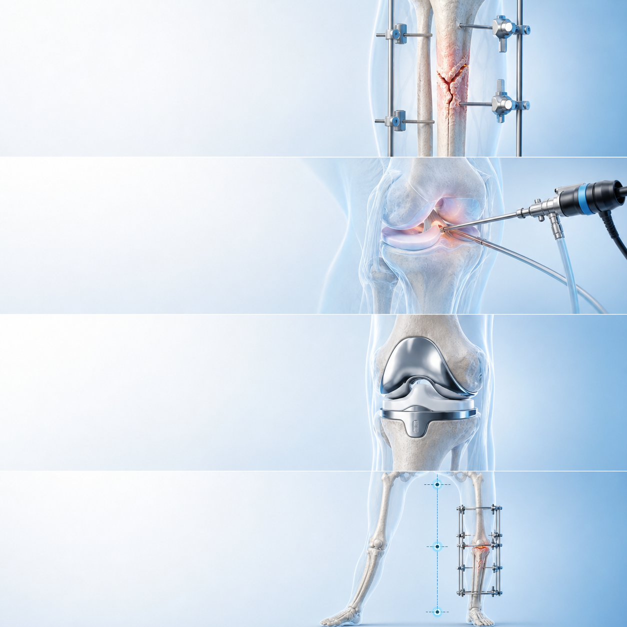
    Can a Meniscus Tear Heal Without Surgery — medically relevant orthopaedic illustration

    Short answer

    Some meniscus tears settle with non-surgical care, while selected tears have limited healing potential or persistent mechanical symptom pattern. Blood supply, location, pattern, associated injury and patient goals influence the decision.

    This guide is provided by Dr Shaharil Orthopedic Specialist Clinic Seremban to help patients understand the clinical review and decision process before an individual consultation.

    Key takeaways

    Main pointSome meniscus tears settle with non-surgical care, while selected tears have limited healing potential or persistent mechanical symptom pattern. Blood supply, location, pattern, associated injury and patient goals influence the decision.
    Useful cluesTrack swelling, locking, instability and response to rehabilitation.
    Clinical reviewThe orthopaedic clinician matches symptom pattern and examination with imaging rather than treating the report alone.
    Possible pathwayOptions may include activity modification, rehabilitation, monitoring, repair or trimming in selected cases.
    Important limitNot all tears can biologically heal, but lack of healing on MRI does not automatically require surgery if symptom pattern are manageable.

    For the complete specialist pathway, symptom pattern assessed and appointment information, read about meniscus management clinical review in Seremban.

    Define the injury or procedure question clearly

    Some meniscus tears settle with non-surgical care, while selected tears have limited healing potential or persistent mechanical symptom pattern. Blood supply, location, pattern, associated injury and patient goals influence the decision. Track swelling, locking, instability and response to rehabilitation.

    Words such as tear, keyhole, instability and sports injury cover very different problems. The useful starting point is the mechanism, exact symptom, functional loss and what has already been tried.

    Match symptom pattern with examination

    The orthopaedic clinician matches symptom pattern and examination with imaging rather than treating the report alone. Associated ligament, cartilage, bone, tendon and kneecap problems may change next-step approach.

    Clinical tests have limitations. They are interpreted together and compared with the movements that matter for work, exercise and sport.

    Use imaging for a reason

    X-rays can show bone, alignment and arthritis. MRI may help with selected soft-tissue questions, but findings such as degenerative meniscus change can occur without being the main cause of pain.

    Appropriate imaging is most useful when it confirms a suspected treatable problem or changes rehabilitation, surgical planning or the need for another pathway.

    Start with the least invasive suitable pathway

    Options may include activity modification, rehabilitation, monitoring, repair or trimming in selected cases. A structured rehabilitation trial should have clear goals, appropriate progression and a review point.

    Surgery is not automatically the faster choice. Recovery after repair, reconstruction or arthroscopy still requires tissue healing and rehabilitation, and restrictions depend on what was done.

    When a procedure may be reasonable

    A procedure is considered when there is a defined diagnosis, symptom pattern and function match it, reasonable alternatives have been discussed and expected benefit outweighs risk.

    For meniscus problems, tear pattern, blood supply, age of injury, tissue quality and associated damage can affect whether repair, trimming or non-surgical care is discussed.

    Plan return to activity with criteria

    Return is safer when movement, strength, balance, confidence and sport-specific control have recovered adequately. Time is one input, not the only clearance test.

    Progress training load gradually and monitor the response during the following day. Recurrent swelling, instability or loss of movement means next-step approach needs reassessment rather than harder training.

    Keep expectations and limitations visible

    Not all tears can biologically heal, but lack of healing on MRI does not automatically require surgery if symptom pattern are manageable. Small incisions do not remove anaesthetic, infection, blood-clot, stiffness or persistent-symptom risks.

    This choice should explain what the chosen management cannot fix. Arthritis, referred pain and deconditioning may require separate management even when a focal injury is treated.

    Know when to seek help

    The practical objective of this process is to connect the clinical findings with the movement, work, family responsibilities or activity that matters to you. A next-step approach should state what to do now, how long to try it and what would trigger review.

    After an injury or procedure, worsening pain, a locked knee, fever, wound drainage, calf swelling, chest pain, breathlessness or new numbness requires timely medical advice.

    Separate return to participation from full performance

    Returning to modified training, completing a full practice and competing at previous intensity are different milestones. For meniscus tear without surgery, define the cutting, landing, running, kneeling, lifting or endurance demands that the knee must tolerate, then rebuild them in a controlled order.

    Monitor the immediate response and the next twenty-four hours. Pain, swelling, loss of movement, instability or reduced control after a progression may mean the load advanced too quickly or the diagnosis needs review. Confidence matters, but confidence should grow alongside strength, movement quality and tissue healing rather than replace them.

    How to use the consultation well

    Bring the questions that would change your decision, not only a request for a scan or procedure. Ask what diagnosis is most likely, what else is being considered, which finding supports next-step approach and what improvement would count as meaningful. For meniscus tear without surgery, it is also helpful to ask what you can safely continue while the clinical review or management is in progress.

    Before leaving, confirm the review point and the safety net. You need to know who will explain any test result, how long the agreed management deserves, which activities should be modified and which new symptom pattern need earlier contact. Write next-step approach down or ask for it in a form you can understand; uncertainty is easier to manage when the next decision is explicit.

    What a responsible plan should contain

    A clinical review-led plan for meniscus tear without surgery should name the working diagnosis, acknowledge important uncertainty and connect each recommendation with a purpose. It should explain expected benefit, relevant harms, alternatives, practical demands and what may happen if management is delayed or declined. When a test is suggested, ask which question it is meant to answer and how each possible result would change the next step.

    Next-step approach should also respect individual health, work, caregiving, financial and activity circumstances without turning those factors into guarantees. Confirm the review date, the measures of progress and the warning signs that override the routine timeline. Clear safety-netting protects patients from waiting too long when the pattern changes, while a defined follow-up prevents unnecessary escalation when recovery is proceeding appropriately.

    What clinical guidance supports

    The AAOS OrthoInfo — Knee Arthroscopy describes arthroscopy as a camera-based procedure for selected joint problems rather than a diagnosis in itself.

    The AAOS OrthoInfo — Meniscus Tears and AAOS — Acute Isolated Meniscal Pathology Guideline show why tear pattern, symptom pattern, repairability and patient factors matter in meniscus decisions.

    NICE — Arthroscopic procedures in osteoarthritis advises against arthroscopic lavage or debridement for osteoarthritis pain alone, while AAOS OrthoInfo — Knee Conditioning Program supports progressive conditioning when appropriate.
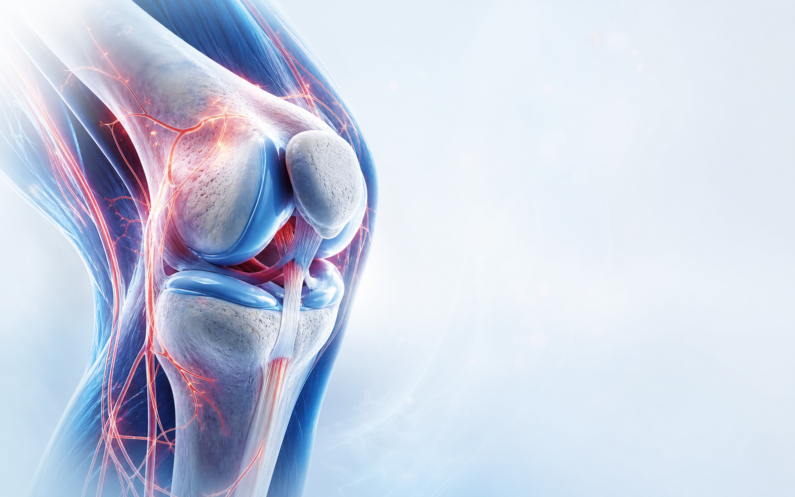
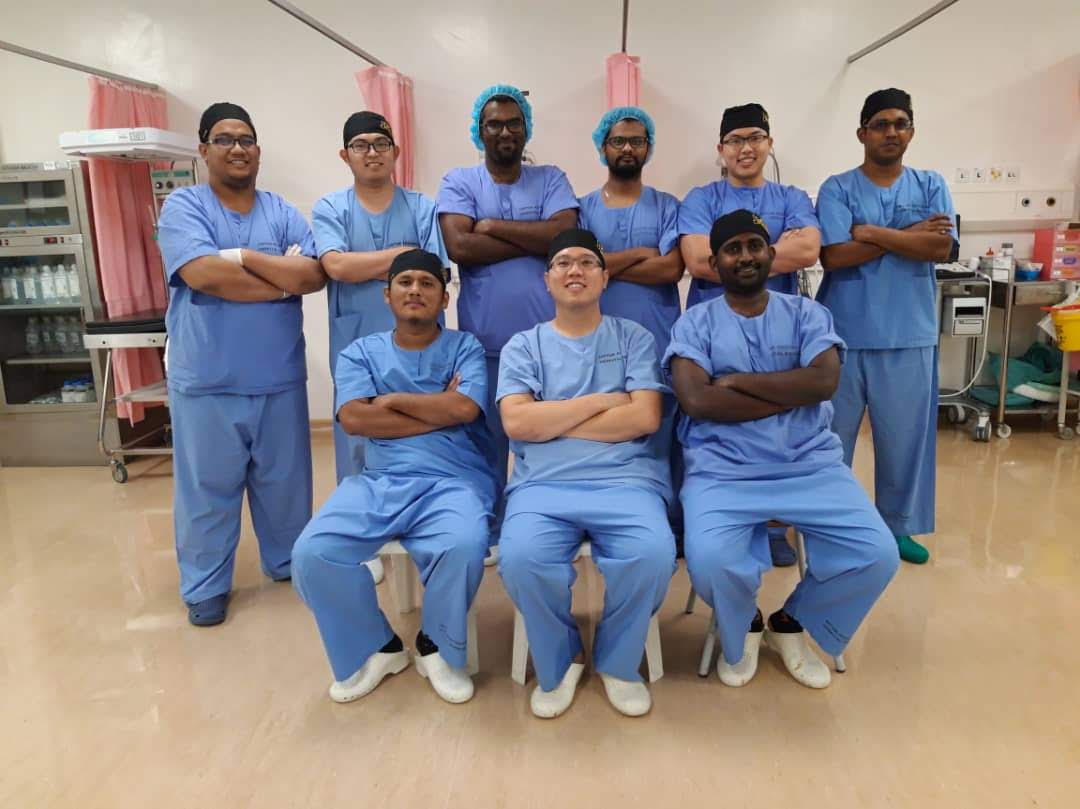
    Clinical orthopaedic illustration relevant to meniscus tear without surgery
    Clinical review connects symptom pattern, examination and appropriate imaging rather than relying on one sign alone.
    Dr Shaharil and orthopaedic clinical team in Seremban
    A clear plan should explain the likely diagnosis, alternatives, limitations, follow-up and urgent warning signs.

    When to seek urgent care

    Prompt review is needed for a knee that remains physically locked or deteriorates after injury.

    Frequently asked questions

    Can meniscus tear without surgery be diagnosed from this symptom alone?

    No. Not all tears can biologically heal, but lack of healing on MRI does not automatically require surgery if symptom pattern are manageable. A focused history and examination are needed before choosing management.

    What should make me seek help sooner?

    Prompt review is needed for a knee that remains physically locked or deteriorates after injury.

    How long should improvement take?

    There is no universal deadline. The diagnosis, severity, health, management and functional demands affect progress; agree on a review point rather than waiting indefinitely.

    Do I need an MRI?

    Not automatically. Appropriate imaging depends on the history, examination, previous results and whether the answer could change management.

    Should I stop all activity?

    Avoid unsafe or sharply aggravating movement, but complete rest is not always helpful. The safe level depends on the diagnosis and severity.

    Can I take pain medicine?

    Ask a doctor or pharmacist what is safe for you, especially with kidney, stomach, liver or heart conditions, pregnancy, blood thinners or other medicines.

    Does seeing an orthopaedic surgeon mean surgery?

    No. Orthopaedic clinical review includes diagnosis, non-surgical planning, monitoring and referral as well as surgery when appropriate.

    What should I bring to the appointment?

    Bring a medicine and allergy list, existing images and reports, relevant management notes and a short timeline of symptom pattern or injury.

    Conclusion

    Some meniscus tears settle with non-surgical care, while selected tears have limited healing potential or persistent mechanical symptom pattern. Blood supply, location, pattern, associated injury and patient goals influence the decision. The safest next step is a proportionate clinical review that connects the symptom or injury with your health, goals and the evidence. A useful plan should also say what would prompt earlier review.

    Medical references

    1. AAOS OrthoInfo — Knee Arthroscopy
    2. AAOS OrthoInfo — Meniscus Tears
    3. AAOS — Acute Isolated Meniscal Pathology Guideline
    4. NICE — Arthroscopic procedures in osteoarthritis
    5. AAOS OrthoInfo — Knee Conditioning Program

    Let’s work out what comes next.

    Tell the clinic what happened, what hurts and which movement has become difficult. The team can help arrange an appointment with Dr Shaharil.

    WhatsApp for an appointment

    This information is general and does not replace an individual medical clinical review, diagnosis or management. If symptom pattern are severe, urgent or worsening, seek appropriate medical care promptly.

  • Meniscus Tear Symptoms: What Should Be Assessed?

    Meniscus Tear Symptoms: What Should Be Assessed?

    Written by Dr ShaharilConsultant Orthopaedic & Trauma Surgeon at CMH Specialist Hospital, Seremban. View Dr Shaharil’s profile.

    Patient guide

    A meniscus tear may cause joint-line pain, swelling, catching, locking or reduced movement, but these concerns overlap with arthritis and other injuries. Diagnosis uses the injury story, examination and imaging when appropriate.

    Meniscus Tear Symptoms What Should Be Assessed — medically relevant orthopaedic illustration

    Short answer

    A meniscus tear may cause joint-line pain, swelling, catching, locking or reduced movement, but these concerns overlap with arthritis and other injuries. Diagnosis uses the injury story, examination and imaging when appropriate.

    This guide is provided by Dr Shaharil Orthopedic Specialist Clinic Seremban to help patients understand the evaluation and decision process before an individual consultation.

    Key takeaways

    Main pointA meniscus tear may cause joint-line pain, swelling, catching, locking or reduced movement, but these concerns overlap with arthritis and other injuries. Diagnosis uses the injury story, examination and imaging when appropriate.
    Useful cluesNote twisting, a pop, delayed swelling, joint-line pain and true mechanical locking.
    EvaluationEvaluation checks movement, tenderness, stability and whether another structure is injured.
    Possible pathwayMany tears do not need immediate surgery; care depends on tear pattern, concerns, age, tissue quality and function.
    Important limitAn MRI finding alone does not prove that the tear causes pain.

    For the complete specialist pathway, concerns assessed and appointment information, read about meniscus and knee arthroscopy evaluation in Seremban.

    Define the injury or procedure question clearly

    A meniscus tear may cause joint-line pain, swelling, catching, locking or reduced movement, but these concerns overlap with arthritis and other injuries. Diagnosis uses the injury story, examination and imaging when appropriate. Note twisting, a pop, delayed swelling, joint-line pain and true mechanical locking.

    Words such as tear, keyhole, instability and sports injury cover very different problems. The useful starting point is the mechanism, exact symptom, functional loss and what has already been tried.

    Match concerns with examination

    Evaluation checks movement, tenderness, stability and whether another structure is injured. Associated ligament, cartilage, bone, tendon and kneecap problems may change pathway.

    Clinical tests have limitations. They are interpreted together and compared with the movements that matter for work, exercise and sport.

    Use imaging for a reason

    X-rays can show bone, alignment and arthritis. MRI may help with selected soft-tissue questions, but findings such as degenerative meniscus change can occur without being the main cause of pain.

    Imaging is most useful when it confirms a suspected treatable problem or changes rehabilitation, surgical planning or the need for another pathway.

    Start with the least invasive suitable pathway

    Many tears do not need immediate surgery; care depends on tear pattern, concerns, age, tissue quality and function. A structured rehabilitation trial should have clear goals, appropriate progression and a review point.

    Surgery is not automatically the faster choice. Recovery after repair, reconstruction or arthroscopy still requires tissue healing and rehabilitation, and restrictions depend on what was done.

    When a procedure may be reasonable

    A procedure is considered when there is a defined diagnosis, concerns and function match it, reasonable alternatives have been discussed and expected benefit outweighs risk.

    For meniscus problems, tear pattern, blood supply, age of injury, tissue quality and associated damage can affect whether repair, trimming or non-surgical care is discussed.

    Plan return to activity with criteria

    Return is safer when movement, strength, balance, confidence and sport-specific control have recovered adequately. Time is one input, not the only clearance test.

    Progress training load gradually and monitor the response during the following day. Recurrent swelling, instability or loss of movement means pathway needs reassessment rather than harder training.

    Keep expectations and limitations visible

    An MRI finding alone does not prove that the tear causes pain. Small incisions do not remove anaesthetic, infection, blood-clot, stiffness or persistent-symptom risks.

    The decision should explain what the chosen care cannot fix. Arthritis, referred pain and deconditioning may require separate management even when a focal injury is treated.

    Know when to seek help

    The goal of this process is to connect the clinical findings with the movement, work, family responsibilities or activity that matters to you. A pathway should state what to do now, how long to try it and what would trigger review.

    After an injury or procedure, worsening pain, a locked knee, fever, wound drainage, calf swelling, chest pain, breathlessness or new numbness requires timely medical advice.

    Separate return to participation from full performance

    Returning to modified training, completing a full practice and competing at previous intensity are different milestones. For meniscus tear concerns, define the cutting, landing, running, kneeling, lifting or endurance demands that the knee must tolerate, then rebuild them in a controlled order.

    Monitor the immediate response and the next twenty-four hours. Pain, swelling, loss of movement, instability or reduced control after a progression may mean the load advanced too quickly or the diagnosis needs review. Confidence matters, but confidence should grow alongside strength, movement quality and tissue healing rather than replace them.

    How to use the consultation well

    Bring the questions that would change your decision, not only a request for a scan or procedure. Ask what diagnosis is most likely, what else is being considered, which finding supports pathway and what improvement would count as meaningful. For meniscus tear concerns, it is also helpful to ask what you can safely continue while the evaluation or care is in progress.

    Before leaving, confirm the review point and the safety net. You should know who will explain any test result, how long the agreed care deserves, which activities should be modified and which new concerns need earlier contact. Write pathway down or ask for it in a form you can understand; uncertainty is easier to manage when the next decision is explicit.

    What a responsible plan should contain

    A evaluation-led plan for meniscus tear concerns should name the working diagnosis, acknowledge important uncertainty and connect each recommendation with a purpose. It should explain expected benefit, relevant harms, alternatives, practical demands and what may happen if care is delayed or declined. When a test is suggested, ask which question it is meant to answer and how each possible result would change the next step.

    Pathway should also respect individual health, work, caregiving, financial and activity circumstances without turning those factors into guarantees. Confirm the review date, the measures of progress and the warning signs that override the routine timeline. Clear safety-netting protects patients from waiting too long when the pattern changes, while a defined follow-up prevents unnecessary escalation when recovery is proceeding appropriately.

    What clinical guidance supports

    The AAOS OrthoInfo — Knee Arthroscopy describes arthroscopy as a camera-based procedure for selected joint problems rather than a diagnosis in itself.

    The AAOS OrthoInfo — Meniscus Tears and AAOS — Acute Isolated Meniscal Pathology Guideline show why tear pattern, concerns, repairability and patient factors matter in meniscus decisions.

    NICE — Arthroscopic procedures in osteoarthritis advises against arthroscopic lavage or debridement for osteoarthritis pain alone, while AAOS OrthoInfo — Knee Conditioning Program supports progressive conditioning when appropriate.

    Clinical orthopaedic illustration relevant to meniscus tear symptoms
    Evaluation connects concerns, examination and appropriate imaging rather than relying on one sign alone.
    Dr Shaharil and orthopaedic clinical team in Seremban
    A clear plan should explain the likely diagnosis, alternatives, limitations, follow-up and urgent warning signs.

    When to seek urgent care

    A truly locked knee, inability to bear weight or major swelling after injury needs prompt evaluation.

    Frequently asked questions

    Can meniscus tear concerns be diagnosed from this symptom alone?

    No. An MRI finding alone does not prove that the tear causes pain. A focused history and examination are needed before choosing care.

    What should make me seek help sooner?

    A truly locked knee, inability to bear weight or major swelling after injury needs prompt evaluation.

    How long should improvement take?

    There is no universal deadline. The diagnosis, severity, health, care and functional demands affect progress; agree on a review point rather than waiting indefinitely.

    Do I need an MRI?

    Not automatically. Imaging depends on the history, examination, previous results and whether the answer could change care.

    Should I stop all activity?

    Avoid unsafe or sharply aggravating movement, but complete rest is not always helpful. The safe level depends on the diagnosis and severity.

    Can I take pain medicine?

    Ask a doctor or pharmacist what is safe for you, especially with kidney, stomach, liver or heart conditions, pregnancy, blood thinners or other medicines.

    Does seeing an orthopaedic surgeon mean surgery?

    No. Orthopaedic evaluation includes diagnosis, non-surgical planning, monitoring and referral as well as surgery when appropriate.

    What should I bring to the appointment?

    Bring a medicine and allergy list, existing images and reports, relevant care notes and a short timeline of concerns or injury.

    Conclusion

    A meniscus tear may cause joint-line pain, swelling, catching, locking or reduced movement, but these concerns overlap with arthritis and other injuries. Diagnosis uses the injury story, examination and imaging when appropriate. The safest next step is a proportionate evaluation that connects the symptom or injury with your health, goals and the evidence. A useful plan should also say what would prompt earlier review.

    Medical references

    1. AAOS OrthoInfo — Knee Arthroscopy
    2. AAOS OrthoInfo — Meniscus Tears
    3. AAOS — Acute Isolated Meniscal Pathology Guideline
    4. NICE — Arthroscopic procedures in osteoarthritis
    5. AAOS OrthoInfo — Knee Conditioning Program

    Let’s work out what comes next.

    Tell the clinic what happened, what hurts and which movement has become difficult. The team can help arrange an appointment with Dr Shaharil.

    WhatsApp for an appointment

    This information is general and does not replace an individual medical evaluation, diagnosis or care. If concerns are severe, urgent or worsening, seek appropriate medical care promptly.
  • Knee Locking, Catching or Giving Way: What Can It Mean?

    Knee Locking, Catching or Giving Way: What Can It Mean?

    Written by Dr ShaharilConsultant Orthopaedic & Trauma Surgeon at CMH Specialist Hospital, Seremban. View Dr Shaharil’s profile.

    When the knee no longer feels dependable

    Locking, catching and giving way are different sensations, but all can reduce confidence in the knee. The exact pattern matters because the same words can describe several different problems.

    Detailed knee anatomy illustration for locking, catching and instability symptoms

    Short answer

    Knee locking or catching may relate to meniscus or cartilage problems, arthritis, inflammation or another mechanical issue. Giving way may follow pain, muscle weakness or ligament instability. A knee that becomes truly locked and cannot straighten, or repeatedly collapses, needs prompt assessment.

    This guide is provided by Dr Shaharil Orthopedic Specialist Clinic Seremban to help patients understand the assessment and treatment pathway before an individual consultation.

    Key takeaways

    Define itTrue locking means the knee physically cannot complete movement; stiffness is different.
    Possible causesMeniscal, ligament, cartilage, kneecap, pain-inhibition and other patterns can overlap.
    Prompt reviewAfter trauma, repeated falls, rapid swelling or inability to fully straighten the knee.
    TreatmentArthroscopy is not automatic; examination and appropriate imaging guide the decision.

    What do locking, catching and giving way mean?

    Catching is often described as a brief snag or interruption during movement. Locking may mean momentary stiffness, or it may describe a knee that becomes physically stuck and cannot fully bend or straighten. Giving way is the feeling that the knee cannot support you reliably.

    Try to describe exactly what happens rather than choosing a diagnosis. Note the movement that triggers it, how long it lasts and whether there is pain, swelling, a pop or a recent injury.

    Several structures may be involved

    Meniscus tears can cause pain, stiffness, swelling, catching, locking and a giving-way sensation. Ligament injury may create instability. Arthritis, cartilage changes, loose fragments, kneecap problems, pain-related inhibition or muscle weakness can produce overlapping symptoms.

    These possibilities cannot be separated reliably by symptoms alone. The age of the patient, injury mechanism, movement pattern, examination and appropriate imaging all contribute to the diagnosis.

    When the symptom needs prompt attention

    A knee that is stuck and cannot straighten, repeated falls because the knee collapses, inability to bear weight or major swelling after an injury should be assessed promptly. A hot, red and swollen knee with fever or feeling unwell requires urgent medical care.

    Do not repeatedly twist or force a locked knee. If the leg looks deformed or circulation and feeling are affected after trauma, go to an Emergency Department.

    Assessment comes before choosing a procedure

    The clinician may check movement, joint-line tenderness, swelling, ligament stability, kneecap tracking, muscle control and walking. X-rays may be useful for bone and arthritis; MRI may be considered when a soft-tissue question remains and the result could change management.

    Treatment depends on the cause. Rehabilitation, activity changes and other non-surgical options may be appropriate. Arthroscopy is considered only for suitable conditions after assessment; locking does not automatically mean keyhole surgery is required.

    What clinical guidance supports

    The AAOS guide to meniscus tears lists catching, locking, giving way, swelling and loss of full motion as possible symptoms, but these symptoms do not prove a meniscal tear on their own.

    After acute knee trauma with concerning findings, the ACR imaging criteria generally support radiographs first; MRI may be the next test when X-rays show no fracture but an internal injury remains suspected. The sequence should follow the clinical question rather than start with an automatic MRI.

    Clinical anatomical illustration of the knee joint in motion
    Knee symptoms can come from different structures, so the pattern and examination matter more than one symptom alone.

    Locking, catching and giving way are not the same

    A true locked knee cannot fully bend or straighten because movement is physically blocked. Catching is a brief interruption or click during motion. Giving way may feel like buckling, but it can result from ligament instability, muscle inhibition caused by pain, kneecap tracking or loss of confidence.

    Use concrete examples when describing the symptom: Did the knee stay stuck? For how long? Did you fall? Was there a twist or pop? Did swelling appear quickly? Can you reproduce the event on stairs, turning or rising from a chair?

    How the cause is investigated

    Assessment may include gait, swelling, range of motion, joint-line tenderness, ligament testing, kneecap movement and strength. The hip and neurological function may be checked when symptoms suggest another source.

    Imaging is selected when it can change treatment. X-rays may identify bone, alignment or arthritis findings. MRI may help when a meniscal, ligament, cartilage or occult bone injury is clinically suspected, but its findings still need to match the symptom pattern.

    When arthroscopy may—and may not—be discussed

    Knee arthroscopy may be considered for selected mechanical problems after the diagnosis, severity, alternatives and likely benefit have been assessed. The word ‘locking’ alone is not an indication for surgery, and many symptoms improve through rehabilitation and load management.

    If a procedure is proposed, ask which structure is being treated, what symptom it is expected to improve, the recovery plan, important risks and what happens without surgery. Shared decisions should include the option of non-surgical care where reasonable.

    How I turn uncertainty into a staged plan

    I usually organise the next step around three questions: what is safe to do now, what missing information would genuinely change treatment, and what development would make us revise the plan. The first step may be activity adjustment, diagnosis-specific rehabilitation, symptom relief or a targeted test. It should be proportionate to both the level of risk and the amount of function you have lost.

    Every plan also needs a review point. We should agree on the change we hope to see, a reasonable timescale and the findings that would mean the strategy is not enough. This avoids waiting indefinitely without direction and also avoids escalating to an invasive treatment before the diagnosis, alternatives and goals are clear.

    Personal factors that can change the recommendation

    Two people with similar scan findings may reasonably choose different plans. Work demands, caregiving, previous injuries, other health conditions, medicines, sport, sleep, willingness to complete rehabilitation and the movement you need to regain can all change the balance of benefit and burden.

    Tell the clinician about diabetes, heart, kidney or stomach problems, blood-thinning medicine, allergies, infection history, pregnancy where relevant, and previous operations or implants. These details do not automatically rule treatment in or out, but they can change medication safety, test selection, procedural risk, preparation and recovery planning.

    Orthopaedic discussion with Dr Shaharil at CMH Specialist Hospital
    A plan is clearer when benefits, limitations, alternatives and follow-up are discussed together.

    Before your assessment, note

    • Whether the knee is briefly catching or completely stuck.
    • Whether you can fully straighten it after the episode.
    • Any twist, fall, pop or rapid swelling.
    • How often the knee gives way and whether it has caused a fall.
    • Pain location and activities that trigger the symptom.

    When to seek urgent care

    Seek prompt or urgent care when the knee is locked and cannot straighten, you cannot bear weight, the knee is deformed or badly swollen after injury, or it is hot and red while you have fever or feel unwell.

    Frequently asked questions

    Is painless knee clicking a concern?

    Painless clicking can be common. Clicking with pain, swelling, locking or instability is more useful to assess.

    Does locking prove that I have a meniscus tear?

    No. Meniscus injury is one possibility, but arthritis and other mechanical or inflammatory problems can cause similar symptoms.

    Will a locked knee need arthroscopy?

    Not automatically. Treatment depends on whether there is a true mechanical block, the underlying cause, severity and the results of assessment.

    Can knee locking or giving way be diagnosed from symptoms alone?

    Symptoms are important, but similar symptoms can arise from different structures or conditions. A diagnosis may require a focused examination and, only when it will change management, an appropriate investigation.

    Should I stop all activity until I am assessed?

    Not automatically. Avoid movements that cause sharp pain, repeated giving way or clear worsening, but complete rest can sometimes increase stiffness and weakness. The safe level of activity depends on the cause and severity.

    Can I take pain medicine before the appointment?

    Do not start, stop or change medication solely because of this article. Ask a doctor or pharmacist what is safe for you, especially if you have kidney, stomach, liver or heart problems, take blood thinners, are pregnant, or use other medicines.

    What if my scan looks normal but I still have pain?

    A scan is one part of assessment. Some painful problems are diagnosed mainly from the history and examination, and some scan findings do not explain the symptoms. Persistent functional difficulty still deserves a clinical review.

    What should I monitor before follow-up?

    Note pain triggers, swelling, sleep disturbance, walking tolerance, work or sport limitations, episodes of locking or giving way, and whether the agreed plan is helping. This gives the follow-up visit useful information.

    Conclusion

    Mechanical knee symptoms deserve precise language and a focused examination. The aim is to distinguish a brief painful catch from a true block or unstable knee, then choose imaging and treatment in proportion to the findings.

    Medical references

    1. AAOS OrthoInfo — Meniscus Tears
    2. NHS — Knee Pain
    3. American College of Radiology — Acute Trauma to the Knee
    4. American College of Radiology — Appropriateness Criteria for Chronic Knee Pain
    5. NICE — Shared Decision Making

    About Dr Shaharil

    Dr Shaharil is a Consultant Orthopaedic & Trauma Surgeon practising at CMH Specialist Hospital in Seremban. Learn more about Dr Shaharil.

    Let’s begin with a clear conversation.

    Tell the clinic what hurts, when it began and what movement has become difficult. The team can help arrange an appointment with Dr Shaharil.

    WhatsApp for an appointment

    This information is general and does not replace an individual medical assessment, diagnosis or treatment. If symptoms are severe or worsening, seek appropriate medical care promptly.
  • Pain at the Front of the Knee: Common Patterns Explained

    Pain at the Front of the Knee: Common Patterns Explained

    Written by Dr ShaharilConsultant Orthopaedic & Trauma Surgeon at CMH Specialist Hospital, Seremban. View Dr Shaharil’s profile.

    Patient guide

    Front-of-knee pain may arise from the kneecap joint, tendons, surrounding soft tissues or referred loading patterns. Activities such as stairs, squatting, running and prolonged sitting help shape the specialist evaluation.

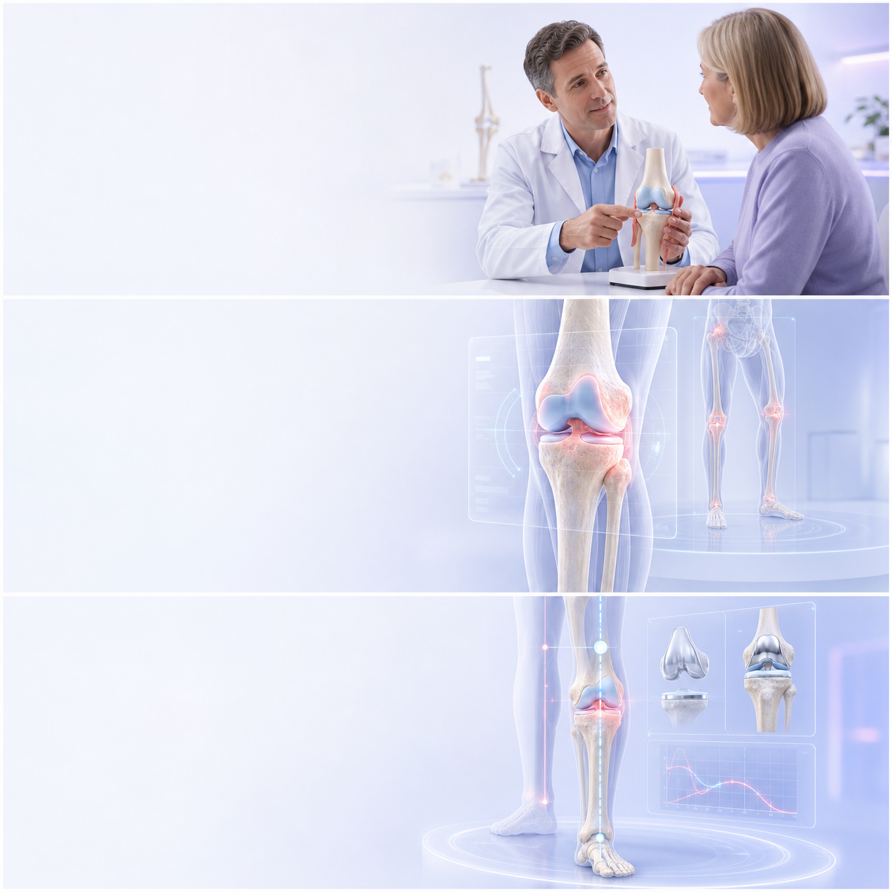
    Pain at the Front of the Knee Common Patterns Explained — medically relevant orthopaedic illustration

    Short answer

    Front-of-knee pain may arise from the kneecap joint, tendons, surrounding soft tissues or referred loading patterns. Activities such as stairs, squatting, running and prolonged sitting help shape the specialist evaluation.

    This guide is provided by Dr Shaharil Orthopedic Specialist Clinic Seremban to help patients understand the specialist evaluation and decision process before an individual consultation.

    Key takeaways

    Main pointFront-of-knee pain may arise from the kneecap joint, tendons, surrounding soft tissues or referred loading patterns. Activities such as stairs, squatting, running and prolonged sitting help shape the specialist evaluation.
    Useful cluesNote whether pain is above, below or around the kneecap, and whether there is clicking, swelling or a recent increase in training.
    Specialist evaluationSpecialist evaluation may include kneecap tracking, tendon tenderness, hip and thigh control, flexibility and functional movements.
    Possible pathwayA graded rehabilitation plan is often important; imaging is reserved for a clear question or a pattern that is not improving as expected.
    Important limitFront knee pain is not one diagnosis and cannot be treated safely by copying a generic exercise list without specialist evaluation.

    For the complete specialist pathway, clinical features assessed and appointment information, read about front knee pain specialist evaluation in Seremban.

    Start with the exact pattern, not a guessed diagnosis

    Front-of-knee pain may arise from the kneecap joint, tendons, surrounding soft tissues or referred loading patterns. Activities such as stairs, squatting, running and prolonged sitting help shape the specialist evaluation. Note whether pain is above, below or around the kneecap, and whether there is clicking, swelling or a recent increase in training.

    The pattern over time matters as much as the pain score. Note what you can no longer do, what remains possible and whether the symptom is improving, stable or gradually becoming more restrictive.

    Why one symptom can have several causes

    Knee clinical features overlap because bone, cartilage, meniscus, ligaments, tendons, bursae, muscles and referred pain can produce similar experiences. Age and activity can shift probabilities, but they do not replace an examination.

    Avoid naming the condition from one movement or an online checklist. A useful description is specific: where it hurts, which direction or load triggers it, what happens afterwards and whether swelling, stiffness, locking or instability accompanies it.

    What to record before the appointment

    Note whether pain is above, below or around the kneecap, and whether there is clicking, swelling or a recent increase in training. A short dated note or photograph of intermittent swelling can be more useful than trying to remember every detail in clinic.

    Include prior injuries, operations, medicines, exercise changes and care pathway already tried. Explain the effect on walking, stairs, prayer positions, sleep, driving, work or sport so care strategy can address real function.

    What a focused specialist evaluation may include

    Specialist evaluation may include kneecap tracking, tendon tenderness, hip and thigh control, flexibility and functional movements. The hip, ankle, back, circulation or nerves may also be considered if the symptom pattern points beyond the knee.

    No single manoeuvre should be treated as a perfect answer. Findings are combined with the history, and their meaning is checked against the tasks that reproduce your clinical features.

    When imaging may add useful information

    X-rays can answer many questions about bone, alignment and arthritis. MRI may be useful for selected soft-tissue questions, but it is not automatically the first or best test for every painful knee.

    A scan is most valuable when the result could change management. Incidental changes are common, so images and reports must be matched to the side, location, timing and examination rather than treated as a diagnosis on their own.

    Care pathway is matched to the cause and goal

    A graded rehabilitation plan is often important; imaging is reserved for a clear question or a pattern that is not improving as expected. Education and a safe progressive plan can be active care pathway even when no procedure is recommended.

    Medication and injections have individual risks and indications. Do not start, stop or change prescribed care pathway from general information; discuss what is safe with the reviewing specialist responsible for your care.

    What this symptom cannot tell you

    Front knee pain is not one diagnosis and cannot be treated safely by copying a generic exercise list without specialist evaluation. Similar clinical features can lead to different plans because severity, diagnosis, health, previous response and goals differ.

    A sensible plan explains uncertainty instead of pretending it does not exist. It should also define when improvement is expected and when persistent or worsening clinical features deserve reassessment.

    Plan the next review point

    The intended outcome of this process is to connect the clinical findings with the movement, work, family responsibilities or activity that matters to you. A care strategy should state what to do now, how long to try it and what would trigger review.

    If progress stalls, the next review may reconsider the diagnosis, exercise dose, adherence, imaging question or need for another opinion. Reassessment is part of good care, not evidence that the first plan failed.

    A practical one-week symptom record

    For seven ordinary days, note the first activity that produces front knee pain, the location, whether the symptom warms up or accumulates, and how the knee feels later that day and the following morning. Record swelling, stiffness, catching or giving way separately rather than combining everything into one pain score.

    Keep the record short enough to use. A few precise entries—such as walking distance, stair direction, sitting duration or number of instability episodes—are more clinically useful than a long diary of every sensation. The intended outcome of this process is not to prove that something is wrong; it is to reveal a repeatable pattern and show how function is changing.

    How to use the consultation well

    Bring the questions that would change your decision, not only a request for a scan or procedure. Ask what diagnosis is most likely, what else is being considered, which finding supports care strategy and what improvement would count as meaningful. For front knee pain, it is also helpful to ask what you can safely continue while the specialist evaluation or care pathway is in progress.

    Before leaving, confirm the review point and the safety net. Patients should know who will explain any test result, how long the agreed care pathway deserves, which activities should be modified and which new clinical features need earlier contact. Write care strategy down or ask for it in a form you can understand; uncertainty is easier to manage when the next decision is explicit.

    What a responsible plan should contain

    A specialist evaluation-led plan for front knee pain should name the working diagnosis, acknowledge important uncertainty and connect each recommendation with a purpose. It should explain expected benefit, relevant harms, alternatives, practical demands and what may happen if care pathway is delayed or declined. When a test is suggested, ask which question it is meant to answer and how each possible result would change the next step.

    Care strategy should also respect individual health, work, caregiving, financial and activity circumstances without turning those factors into guarantees. Confirm the review date, the measures of progress and the warning signs that override the routine timeline. Clear safety-netting protects patients from waiting too long when the pattern changes, while a defined follow-up prevents unnecessary escalation when recovery is proceeding appropriately.

    What clinical guidance supports

    The NHS — Knee pain advises specialist evaluation when knee pain persists, affects function, or occurs with locking, giving way, marked swelling or inability to bear weight.

    NICE — Osteoarthritis: specialist evaluation and management emphasises clinical features and physical function in specialist evaluation and recommends core non-surgical care rather than using imaging severity alone.

    The American College of Radiology — Chronic Knee Pain supports selecting imaging according to the clinical scenario, while the AAOS OrthoInfo — Meniscus Tears illustrates why pain, swelling, catching and giving way can overlap across conditions.

    Clinical orthopaedic illustration relevant to front knee pain
    Specialist evaluation connects clinical features, examination and appropriate imaging rather than relying on one sign alone.
    Dr Shaharil and orthopaedic clinical team in Seremban
    A clear plan should explain the likely diagnosis, alternatives, limitations, follow-up and urgent warning signs.

    When to seek urgent care

    Seek urgent review after trauma if the kneecap looks displaced, the knee cannot straighten or weight-bearing is impossible.

    Frequently asked questions

    Can front knee pain be diagnosed from this symptom alone?

    No. Front knee pain is not one diagnosis and cannot be treated safely by copying a generic exercise list without specialist evaluation. A focused history and examination are needed before choosing care pathway.

    What should make me seek help sooner?

    Seek urgent review after trauma if the kneecap looks displaced, the knee cannot straighten or weight-bearing is impossible.

    How long should improvement take?

    There is no universal deadline. The diagnosis, severity, health, care pathway and functional demands affect progress; agree on a review point rather than waiting indefinitely.

    Do I need an MRI?

    Not automatically. Radiological evaluation depends on the history, examination, previous results and whether the answer could change care pathway.

    Should I stop all activity?

    Avoid unsafe or sharply aggravating movement, but complete rest is not always helpful. The safe level depends on the diagnosis and severity.

    Can I take pain medicine?

    Ask a doctor or pharmacist what is safe for you, especially with kidney, stomach, liver or heart conditions, pregnancy, blood thinners or other medicines.

    Does seeing an orthopaedic surgeon mean surgery?

    No. Orthopaedic specialist evaluation includes diagnosis, non-surgical planning, monitoring and referral as well as surgery when appropriate.

    What should I bring to the appointment?

    Bring a medicine and allergy list, existing images and reports, relevant care pathway notes and a short timeline of clinical features or injury.

    Conclusion

    Front-of-knee pain may arise from the kneecap joint, tendons, surrounding soft tissues or referred loading patterns. Activities such as stairs, squatting, running and prolonged sitting help shape the specialist evaluation. The safest next step is a proportionate specialist evaluation that connects the symptom or injury with your health, goals and the evidence. A useful plan should also say what would prompt earlier review.

    Medical references

    1. NHS — Knee pain
    2. NICE — Osteoarthritis: specialist evaluation and management
    3. American College of Radiology — Chronic Knee Pain
    4. AAOS OrthoInfo — Meniscus Tears
    5. AAOS OrthoInfo — Knee Conditioning Program

    Let’s work out what comes next.

    Tell the clinic what happened, what hurts and which movement has become difficult. The team can help arrange an appointment with Dr Shaharil.

    WhatsApp for an appointment

    This information is general and does not replace an individual medical specialist evaluation, diagnosis or care pathway. If clinical features are severe, urgent or worsening, seek appropriate medical care promptly.

  • Why Does My Knee Hurt When Climbing Stairs?

    Why Does My Knee Hurt When Climbing Stairs?

    Written by Dr ShaharilConsultant Orthopaedic & Trauma Surgeon at CMH Specialist Hospital, Seremban. View Dr Shaharil’s profile.

    Patient guide

    Knee pain on stairs often reflects how the kneecap, joint surfaces, tendons and muscles manage load. The direction of travel, pain location and presence of swelling or instability help determine what should be assessed.

    Why Does My Knee Hurt When Climbing Stairs — medically relevant orthopaedic illustration

    Short answer

    Knee pain on stairs often reflects how the kneecap, joint surfaces, tendons and muscles manage load. The direction of travel, pain location and presence of swelling or instability help determine what should be assessed.

    This guide is provided by Dr Shaharil Orthopedic Specialist Clinic Seremban to help patients understand the evaluation and decision process before an individual consultation.

    Key takeaways

    Main pointKnee pain on stairs often reflects how the kneecap, joint surfaces, tendons and muscles manage load. The direction of travel, pain location and presence of swelling or instability help determine what should be assessed.
    Useful cluesPain may be worse going down, going up, after sitting or only when carrying weight. Note whether it is in front, inside, outside or deep within the knee.
    EvaluationEvaluation considers kneecap tracking, joint-line tenderness, movement, strength, alignment and walking rather than using the stair symptom as a diagnosis.
    Possible pathwayA pathway may include load adjustment, rehabilitation, medication review or targeted imaging when it can change management.
    Important limitStair pain does not by itself prove arthritis, cartilage loss or a need for surgery.

    For the complete specialist pathway, concerns assessed and appointment information, read about knee pain evaluation in Seremban.

    Start with the exact pattern, not a guessed diagnosis

    Knee pain on stairs often reflects how the kneecap, joint surfaces, tendons and muscles manage load. The direction of travel, pain location and presence of swelling or instability help determine what should be assessed. Pain may be worse going down, going up, after sitting or only when carrying weight. Note whether it is in front, inside, outside or deep within the knee.

    The pattern over time matters as much as the pain score. Note what you can no longer do, what remains possible and whether the symptom is improving, stable or gradually becoming more restrictive.

    Why one symptom can have several causes

    Knee concerns overlap because bone, cartilage, meniscus, ligaments, tendons, bursae, muscles and referred pain can produce similar experiences. Age and activity can shift probabilities, but they do not replace an examination.

    Avoid naming the condition from one movement or an online checklist. A useful description is specific: where it hurts, which direction or load triggers it, what happens afterwards and whether swelling, stiffness, locking or instability accompanies it.

    What to record before the appointment

    Pain may be worse going down, going up, after sitting or only when carrying weight. Note whether it is in front, inside, outside or deep within the knee. A short dated note or photograph of intermittent swelling can be more useful than trying to remember every detail in clinic.

    Include prior injuries, operations, medicines, exercise changes and care already tried. Explain the effect on walking, stairs, prayer positions, sleep, driving, work or sport so pathway can address real function.

    What a focused evaluation may include

    Evaluation considers kneecap tracking, joint-line tenderness, movement, strength, alignment and walking rather than using the stair symptom as a diagnosis. The hip, ankle, back, circulation or nerves may also be considered if the symptom pattern points beyond the knee.

    No single manoeuvre should be treated as a perfect answer. Findings are combined with the history, and their meaning is checked against the tasks that reproduce your concerns.

    When imaging may add useful information

    X-rays can answer many questions about bone, alignment and arthritis. MRI may be useful for selected soft-tissue questions, but it is not automatically the first or best test for every painful knee.

    A scan is most valuable when the result could change management. Incidental changes are common, so images and reports must be matched to the side, location, timing and examination rather than treated as a diagnosis on their own.

    Care is matched to the cause and goal

    A pathway may include load adjustment, rehabilitation, medication review or targeted imaging when it can change management. Education and a safe progressive plan can be active care even when no procedure is recommended.

    Medication and injections have individual risks and indications. Do not start, stop or change prescribed care from general information; discuss what is safe with the specialist responsible for your care.

    What this symptom cannot tell you

    Stair pain does not by itself prove arthritis, cartilage loss or a need for surgery. Similar concerns can lead to different plans because severity, diagnosis, health, previous response and goals differ.

    A sensible plan explains uncertainty instead of pretending it does not exist. It should also define when improvement is expected and when persistent or worsening concerns deserve reassessment.

    Plan the next review point

    The goal of this process is to connect the clinical findings with the movement, work, family responsibilities or activity that matters to you. A pathway should state what to do now, how long to try it and what would trigger review.

    If progress stalls, the next review may reconsider the diagnosis, exercise dose, adherence, imaging question or need for another opinion. Reassessment is part of good care, not evidence that the first plan failed.

    A practical one-week symptom record

    For seven ordinary days, note the first activity that produces knee pain climbing stairs, the location, whether the symptom warms up or accumulates, and how the knee feels later that day and the following morning. Record swelling, stiffness, catching or giving way separately rather than combining everything into one pain score.

    Keep the record short enough to use. A few precise entries—such as walking distance, stair direction, sitting duration or number of instability episodes—are more clinically useful than a long diary of every sensation. The goal of this process is not to prove that something is wrong; it is to reveal a repeatable pattern and show how function is changing.

    How to use the consultation well

    Bring the questions that would change your decision, not only a request for a scan or procedure. Ask what diagnosis is most likely, what else is being considered, which finding supports pathway and what improvement would count as meaningful. For knee pain climbing stairs, it is also helpful to ask what you can safely continue while the evaluation or care is in progress.

    Before leaving, confirm the review point and the safety net. You should know who will explain any test result, how long the agreed care deserves, which activities should be modified and which new concerns need earlier contact. Write pathway down or ask for it in a form you can understand; uncertainty is easier to manage when the next decision is explicit.

    What a responsible plan should contain

    A evaluation-led plan for knee pain climbing stairs should name the working diagnosis, acknowledge important uncertainty and connect each recommendation with a purpose. It should explain expected benefit, relevant harms, alternatives, practical demands and what may happen if care is delayed or declined. When a test is suggested, ask which question it is meant to answer and how each possible result would change the next step.

    Pathway should also respect individual health, work, caregiving, financial and activity circumstances without turning those factors into guarantees. Confirm the review date, the measures of progress and the warning signs that override the routine timeline. Clear safety-netting protects patients from waiting too long when the pattern changes, while a defined follow-up prevents unnecessary escalation when recovery is proceeding appropriately.

    What clinical guidance supports

    The NHS — Knee pain advises evaluation when knee pain persists, affects function, or occurs with locking, giving way, marked swelling or inability to bear weight.

    NICE — Osteoarthritis: evaluation and management emphasises concerns and physical function in evaluation and recommends core non-surgical care rather than using imaging severity alone.

    The American College of Radiology — Chronic Knee Pain supports selecting imaging according to the clinical scenario, while the AAOS OrthoInfo — Meniscus Tears illustrates why pain, swelling, catching and giving way can overlap across conditions.

    Clinical orthopaedic illustration relevant to knee pain climbing stairs
    Evaluation connects concerns, examination and appropriate imaging rather than relying on one sign alone.
    Dr Shaharil and orthopaedic clinical team in Seremban
    A clear plan should explain the likely diagnosis, alternatives, limitations, follow-up and urgent warning signs.

    When to seek urgent care

    Seek urgent care if a recent injury caused deformity, inability to bear weight, a locked knee or a hot red joint with fever.

    Frequently asked questions

    Can knee pain climbing stairs be diagnosed from this symptom alone?

    No. Stair pain does not by itself prove arthritis, cartilage loss or a need for surgery. A focused history and examination are needed before choosing care.

    What should make me seek help sooner?

    Seek urgent care if a recent injury caused deformity, inability to bear weight, a locked knee or a hot red joint with fever.

    How long should improvement take?

    There is no universal deadline. The diagnosis, severity, health, care and functional demands affect progress; agree on a review point rather than waiting indefinitely.

    Do I need an MRI?

    Not automatically. Imaging depends on the history, examination, previous results and whether the answer could change care.

    Should I stop all activity?

    Avoid unsafe or sharply aggravating movement, but complete rest is not always helpful. The safe level depends on the diagnosis and severity.

    Can I take pain medicine?

    Ask a doctor or pharmacist what is safe for you, especially with kidney, stomach, liver or heart conditions, pregnancy, blood thinners or other medicines.

    Does seeing an orthopaedic surgeon mean surgery?

    No. Orthopaedic evaluation includes diagnosis, non-surgical planning, monitoring and referral as well as surgery when appropriate.

    What should I bring to the appointment?

    Bring a medicine and allergy list, existing images and reports, relevant care notes and a short timeline of concerns or injury.

    Conclusion

    Knee pain on stairs often reflects how the kneecap, joint surfaces, tendons and muscles manage load. The direction of travel, pain location and presence of swelling or instability help determine what should be assessed. The safest next step is a proportionate evaluation that connects the symptom or injury with your health, goals and the evidence. A useful plan should also say what would prompt earlier review.

    Medical references

    1. NHS — Knee pain
    2. NICE — Osteoarthritis: evaluation and management
    3. American College of Radiology — Chronic Knee Pain
    4. AAOS OrthoInfo — Meniscus Tears
    5. AAOS OrthoInfo — Knee Conditioning Program

    Let’s work out what comes next.

    Tell the clinic what happened, what hurts and which movement has become difficult. The team can help arrange an appointment with Dr Shaharil.

    WhatsApp for an appointment

    This information is general and does not replace an individual medical evaluation, diagnosis or care. If concerns are severe, urgent or worsening, seek appropriate medical care promptly.
  • Why Does My Knee Swell After Walking or Exercise?

    Why Does My Knee Swell After Walking or Exercise?

    Written by Dr ShaharilConsultant Orthopaedic & Trauma Surgeon at CMH Specialist Hospital, Seremban. View Dr Shaharil’s profile.

    Swelling is a signal, not a diagnosis

    A knee that becomes puffy or tight after activity is telling you that the joint or surrounding tissues have been irritated. The timing, amount of swelling and other symptoms help determine how quickly it should be assessed.

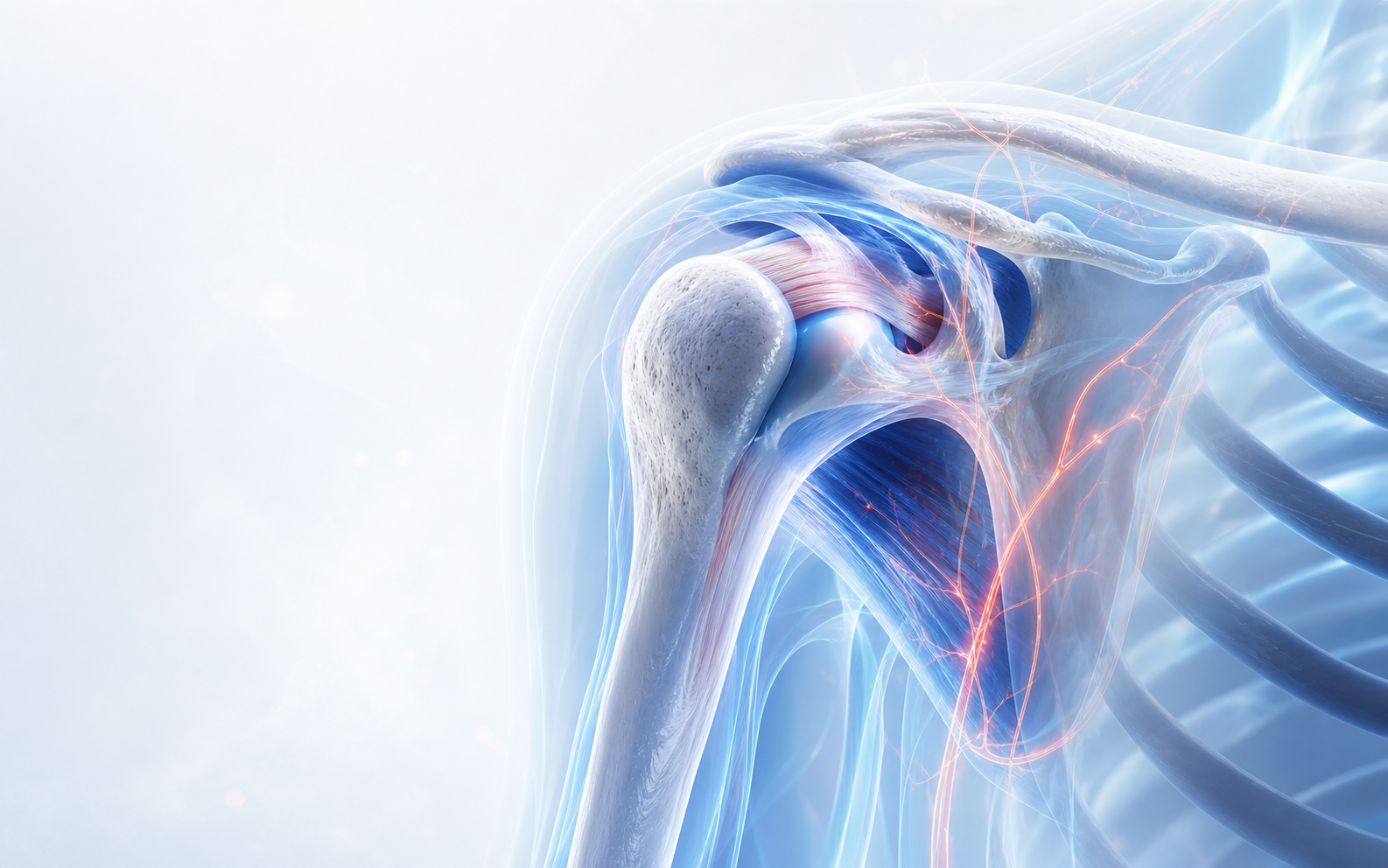
    Anatomical illustration of the knee for an article about swelling after activity

    Short answer

    Knee swelling after walking or exercise may follow overuse, irritation from arthritis, a meniscus or ligament injury, inflammation around the joint or another condition. The cause cannot be confirmed from swelling alone. Recurrent, sudden or severe swelling should be assessed, particularly when movement or weight-bearing is affected.

    This guide is provided by Dr Shaharil Orthopedic Specialist Clinic Seremban to help patients understand the assessment and treatment pathway before an individual consultation.

    Key takeaways

    MeaningSwelling is a response, not a diagnosis; overload, irritation or injury are possible.
    TrackOnset, activity dose, location, heat, stiffness, locking and recovery time.
    AssessmentRecurrent or increasing swelling, instability or loss of movement should be examined.
    UrgentA hot painful swollen knee with fever, major injury or inability to bear weight needs urgent care.

    Notice when the swelling appears

    Swelling that develops immediately after a twist or impact may carry a different meaning from mild puffiness that increases gradually after a long walk. Note whether it begins within minutes, later the same day or the following morning.

    Also notice whether the knee feels warm, stiff or unstable, whether you can straighten it fully and whether the swelling settles between episodes. These details are useful during assessment.

    Common categories—not a self-diagnosis

    Repeated loading can irritate tissues around the knee. Arthritis can also cause activity-related pain, stiffness and swelling. After a twist, symptoms may involve the meniscus, ligaments or cartilage. Bursitis and inflammatory conditions are other possibilities.

    Several problems can feel similar. Age, previous injury, activity pattern, examination and sometimes imaging are needed to distinguish them. Avoid assuming that swelling automatically means worn cartilage or a torn meniscus.

    What you can record before the appointment

    Write down the activity, distance or movement that preceded the swelling. A dated photograph can help if the knee looks normal by the time of the appointment. Mention any locking, giving way, fever, redness, calf swelling or inability to bear weight.

    Describe what you have tried and whether it helped. Do not repeatedly push through activity that causes marked swelling or instability while waiting for assessment.

    How swelling may be assessed

    The clinician may compare both knees, check where the swelling sits, assess movement and stability and look for signs suggesting injury, arthritis or inflammation. Imaging or other tests are used when they can answer a relevant clinical question.

    Treatment depends on the cause. It may involve temporary activity changes, rehabilitation, medication discussion, treatment of an injury or arthritis, or further investigation. An operation is not the automatic response to swelling.

    What clinical guidance supports

    The NHS knee-pain guide lists several patterns associated with swelling and recommends urgent assessment when severe swelling follows injury or when the knee is hot, red and accompanied by fever.

    A meniscal injury is one possible—not automatic—cause of swelling, catching or giving way after a twist, as described by AAOS OrthoInfo. For typical osteoarthritis, NICE recommends basing management on symptoms and function rather than repeatedly scanning the joint.

    Clinical anatomical illustration of the knee joint in motion
    Knee symptoms can come from different structures, so the pattern and examination matter more than one symptom alone.

    Why activity can reveal swelling

    Walking and exercise increase load and joint-fluid movement. A sudden jump in distance, hills, squats, running or sport can irritate tissues that are not yet conditioned for that demand. Arthritis, synovial irritation, meniscal problems and other causes may produce a similar visible response.

    Timing gives clues. Swelling within hours of a twist is different from mild puffiness that appears after a long walk and settles overnight. Note whether the knee feels hot, whether you can fully bend and straighten it, and whether the swelling is inside the joint or in a more localised area.

    What you can do while arranging assessment

    Reduce the activity dose that clearly triggers swelling and use relative rest rather than complete immobility. A wrapped cold pack for a short period may help comfort if safe for your skin and sensation. Do not force deep bending or continue sport through repeated giving way.

    Do not start anti-inflammatory medicines without checking whether they are safe for you. Kidney disease, stomach ulcers, blood thinners, cardiovascular conditions, pregnancy and other medicines can change the risk.

    When recurrent swelling changes the plan

    Swelling that repeatedly returns, lasts longer, appears with locking or instability, or progressively limits motion deserves clinical assessment. The examination can help decide whether rehabilitation, an X-ray, MRI, fluid testing or another step is justified.

    The goal is not simply to remove swelling; it is to identify and manage the reason the knee keeps reacting. Draining fluid or obtaining a scan is not automatically necessary for every swollen knee.

    How I turn uncertainty into a staged plan

    I usually organise the next step around three questions: what is safe to do now, what missing information would genuinely change treatment, and what development would make us revise the plan. The first step may be activity adjustment, diagnosis-specific rehabilitation, symptom relief or a targeted test. It should be proportionate to both the level of risk and the amount of function you have lost.

    Every plan also needs a review point. We should agree on the change we hope to see, a reasonable timescale and the findings that would mean the strategy is not enough. This avoids waiting indefinitely without direction and also avoids escalating to an invasive treatment before the diagnosis, alternatives and goals are clear.

    Personal factors that can change the recommendation

    Two people with similar scan findings may reasonably choose different plans. Work demands, caregiving, previous injuries, other health conditions, medicines, sport, sleep, willingness to complete rehabilitation and the movement you need to regain can all change the balance of benefit and burden.

    Tell the clinician about diabetes, heart, kidney or stomach problems, blood-thinning medicine, allergies, infection history, pregnancy where relevant, and previous operations or implants. These details do not automatically rule treatment in or out, but they can change medication safety, test selection, procedural risk, preparation and recovery planning.

    Dr Shaharil discussing orthopaedic care in a clinical setting
    Bring your questions and existing records so the discussion can focus on a safe, practical next step.

    Useful details to track

    • How quickly the swelling develops after activity.
    • Whether the knee is hot, red, stiff, locked or unstable.
    • Whether you can walk and fully straighten the knee.
    • Which activities trigger it and how long it lasts.
    • Any recent fall, twist, increase in training or previous knee injury.

    When to seek urgent care

    Seek urgent care for severe or rapidly increasing swelling after injury, inability to bear weight, deformity, a locked knee, or a hot red swollen knee with fever or feeling unwell. Significant calf swelling, chest pain or breathlessness also needs urgent medical attention.

    Frequently asked questions

    Is knee swelling after exercise normal?

    Temporary mild symptoms can occur after unusual loading, but recurrent, marked or painful swelling is not something to ignore. It should be assessed if it persists or affects function.

    Should I keep exercising on a swollen knee?

    Avoid forcing activity that causes marked pain, swelling or instability. The appropriate level of movement depends on the cause and severity.

    Does swelling mean I need surgery?

    No. Many causes are treated non-surgically. Treatment depends on the diagnosis and how the symptoms affect you.

    Can knee swelling be diagnosed from symptoms alone?

    Symptoms are important, but similar symptoms can arise from different structures or conditions. A diagnosis may require a focused examination and, only when it will change management, an appropriate investigation.

    Should I stop all activity until I am assessed?

    Not automatically. Avoid movements that cause sharp pain, repeated giving way or clear worsening, but complete rest can sometimes increase stiffness and weakness. The safe level of activity depends on the cause and severity.

    Can I take pain medicine before the appointment?

    Do not start, stop or change medication solely because of this article. Ask a doctor or pharmacist what is safe for you, especially if you have kidney, stomach, liver or heart problems, take blood thinners, are pregnant, or use other medicines.

    What if my scan looks normal but I still have pain?

    A scan is one part of assessment. Some painful problems are diagnosed mainly from the history and examination, and some scan findings do not explain the symptoms. Persistent functional difficulty still deserves a clinical review.

    What should I monitor before follow-up?

    Note pain triggers, swelling, sleep disturbance, walking tolerance, work or sport limitations, episodes of locking or giving way, and whether the agreed plan is helping. This gives the follow-up visit useful information.

    Conclusion

    A knee that swells after activity is telling you that the current load exceeds what the joint can comfortably tolerate—or that another problem needs attention. Track the pattern, adjust the load and seek assessment if swelling is recurrent, increasing or mechanically disruptive.

    Medical references

    1. NHS — Knee Pain
    2. NHS — Joint pain
    3. AAOS OrthoInfo — Meniscus Tears
    4. NICE — Osteoarthritis in over 16s: diagnosis and management
    5. American College of Radiology — Appropriateness Criteria for Chronic Knee Pain

    About Dr Shaharil

    Dr Shaharil is a Consultant Orthopaedic & Trauma Surgeon practising at CMH Specialist Hospital in Seremban. Learn more about Dr Shaharil.

    Let’s begin with a clear conversation.

    Tell the clinic what hurts, when it began and what movement has become difficult. The team can help arrange an appointment with Dr Shaharil.

    WhatsApp for an appointment

    This information is general and does not replace an individual medical assessment, diagnosis or treatment. If symptoms are severe or worsening, seek appropriate medical care promptly.

  • Does Seeing an Orthopaedic Surgeon Always Mean Surgery?

    Does Seeing an Orthopaedic Surgeon Always Mean Surgery?

    Written by Dr ShaharilConsultant Orthopaedic & Trauma Surgeon at CMH Specialist Hospital, Seremban. View Dr Shaharil’s profile.

    Assessment first, treatment second

    The word surgeon can make an appointment sound like a decision has already been made. It has not. Orthopaedic surgeons assess musculoskeletal conditions and use both non-surgical and surgical treatment pathways.

    Orthopaedic consultation illustration representing treatment choices

    Short answer

    No. An orthopaedic surgeon may recommend advice, rehabilitation, activity modification, medication, bracing, monitoring or another non-surgical option. Surgery is considered when the diagnosis, severity, likely benefits, limitations and your goals make it an appropriate choice.

    This guide is provided by Dr Shaharil Orthopedic Specialist Clinic Seremban to help patients understand the assessment and treatment pathway before an individual consultation.

    Key takeaways

    No commitmentAn orthopaedic consultation is an assessment, not consent for surgery.
    OptionsEducation, rehabilitation, activity change, medicines or injections may be considered where suitable.
    SurgeryDiscussed when the diagnosis, symptom burden and expected benefit justify it.
    Your choiceReasonable alternatives, risks, limitations and the option to wait should be explained.

    Orthopaedic care begins with diagnosis

    Bones, joints, ligaments, tendons and muscles can produce similar symptoms for different reasons. Before discussing treatment, the clinician needs to understand the history, examine the affected area and decide whether any investigation is necessary.

    Sometimes the most valuable outcome of the consultation is a clearer diagnosis and a structured non-surgical plan. In other situations, the assessment may show that monitoring, referral to another service or urgent care is more suitable.

    Non-surgical options are part of orthopaedics

    Depending on the condition, non-surgical care may include education, changes to activity, rehabilitation, medication, a brace or cast, weight-management support, injections when clinically appropriate, or follow-up over time. Not every option suits every person.

    The plan should address what you need to do in daily life and what outcome is realistic. A scan finding alone should not force a treatment decision without considering symptoms, function and personal priorities.

    When surgery enters the conversation

    Surgery may be discussed for a significant injury, progressive damage, instability, deformity, symptoms that remain limiting despite appropriate non-surgical care, or another problem where an operation offers a reasonable balance of benefit and risk.

    A recommendation is not the same as pressure. You should understand the purpose of the operation, alternatives, expected recovery, important risks, likely limitations and what may happen if you choose not to proceed at that time.

    The decision should be shared

    Shared decision making combines clinical evidence and professional judgement with your preferences and circumstances. The best option may differ between two people with similar imaging because their symptoms, health, work, support and goals are different.

    Ask for explanations in plain language. It is reasonable to take time, discuss the decision with family, return with more questions or seek another opinion when the situation is not urgent.

    What clinical guidance supports

    The AAOS description of orthopaedic practice includes diagnosis, prevention, rehabilitation and both non-surgical and surgical treatment. Seeing a surgeon therefore does not mean an operation has been chosen.

    The GMC consent principles require meaningful dialogue about benefits, harms, reasonable alternatives and the option to take no action. NICE osteoarthritis guidance likewise places exercise, information and appropriate weight management at the core of care before joint-replacement referral is considered for substantially affected quality of life.

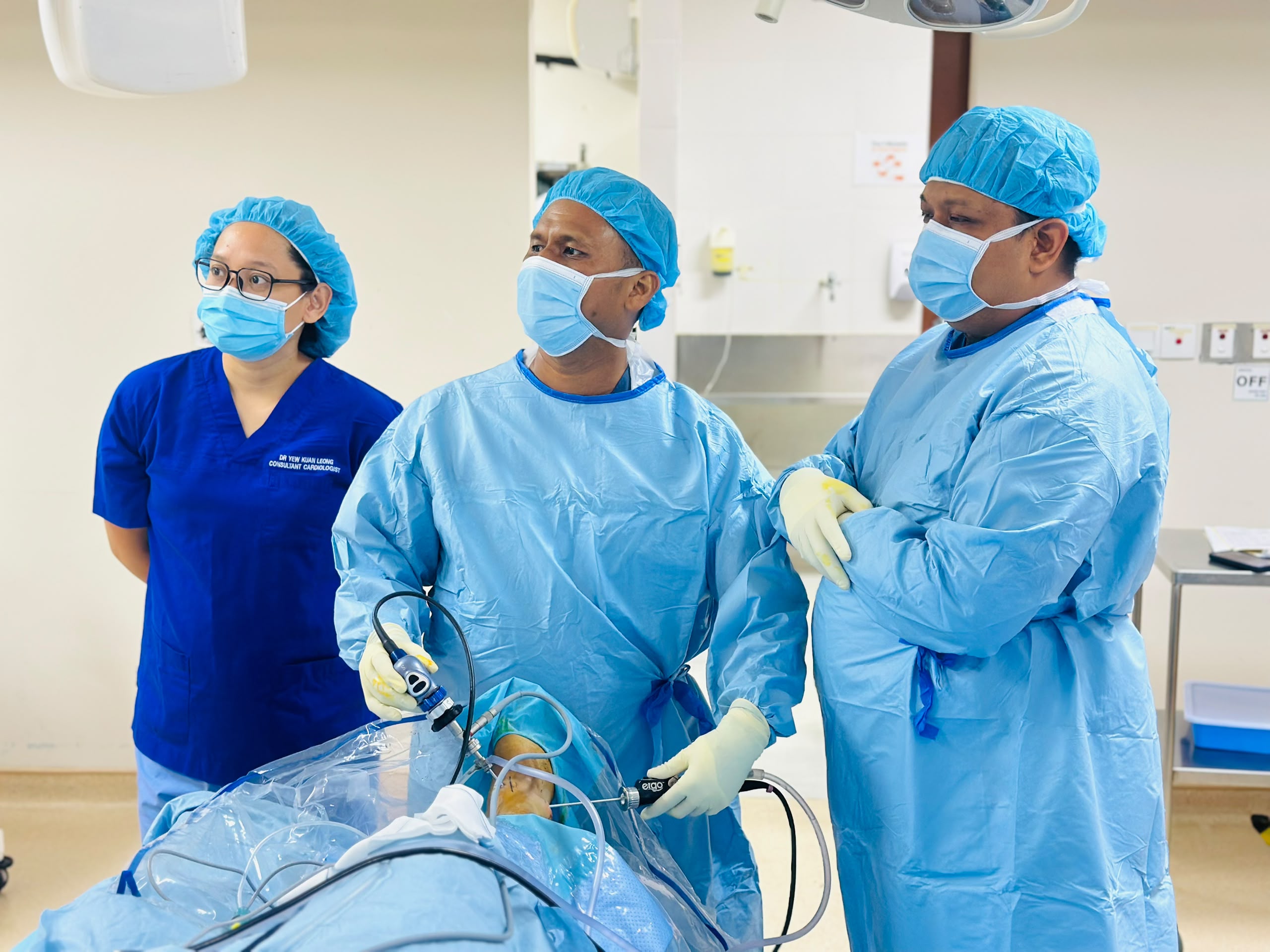
    Dr Shaharil, Consultant Orthopaedic and Trauma Surgeon in Seremban
    A consultation should connect the clinical findings with the movement and daily activities that matter to you.

    What non-surgical care can involve

    A non-surgical plan may combine diagnosis-specific education, activity modification, progressive exercise or physiotherapy, occupational changes, a brace or walking aid, and medication review. Injections may be appropriate for selected conditions but are not a universal solution.

    The plan should state what improvement is realistic, how long to try it and what would count as failure. Non-surgical treatment is active treatment—not dismissal—and it needs the same follow-up discipline as a procedure.

    When surgery enters the discussion

    Surgery is more likely to be discussed when there is a structural problem that can be treated surgically, symptoms materially affect quality of life or safety, and reasonable alternatives are ineffective, unsuitable or unlikely to solve the problem. Urgent injuries are a different pathway from long-standing degenerative pain.

    A surgeon should explain the purpose of the procedure, important risks, expected recovery, rehabilitation demands and the chance that some symptoms may remain. No operation can guarantee a perfect result.

    It is reasonable to pause and ask questions

    Unless treatment is urgent, you can take time to consider the information, involve family and seek clarification. Ask what happens if you wait, which symptoms might progress, and whether a second opinion would be useful.

    Your decision should reflect your health, responsibilities, risk tolerance and goals. A technically possible operation is not automatically the best option for every person.

    How I turn uncertainty into a staged plan

    I usually organise the next step around three questions: what is safe to do now, what missing information would genuinely change treatment, and what development would make us revise the plan. The first step may be activity adjustment, diagnosis-specific rehabilitation, symptom relief or a targeted test. It should be proportionate to both the level of risk and the amount of function you have lost.

    Every plan also needs a review point. We should agree on the change we hope to see, a reasonable timescale and the findings that would mean the strategy is not enough. This avoids waiting indefinitely without direction and also avoids escalating to an invasive treatment before the diagnosis, alternatives and goals are clear.

    Personal factors that can change the recommendation

    Two people with similar scan findings may reasonably choose different plans. Work demands, caregiving, previous injuries, other health conditions, medicines, sport, sleep, willingness to complete rehabilitation and the movement you need to regain can all change the balance of benefit and burden.

    Tell the clinician about diabetes, heart, kidney or stomach problems, blood-thinning medicine, allergies, infection history, pregnancy where relevant, and previous operations or implants. These details do not automatically rule treatment in or out, but they can change medication safety, test selection, procedural risk, preparation and recovery planning.

    Orthopaedic discussion with Dr Shaharil at CMH Specialist Hospital
    A plan is clearer when benefits, limitations, alternatives and follow-up are discussed together.

    Questions worth asking

    • What are the realistic non-surgical options for my condition?
    • What problem would surgery be intended to solve?
    • What are the benefits, limitations and important risks?
    • What could happen if I wait or continue non-surgical treatment?
    • What recovery support would I need?

    When to seek urgent care

    Shared decision making applies to planned care, not emergencies. Major trauma, an open wound over a possible fracture, threatened circulation or sensation, or a hot swollen joint with fever requires urgent assessment.

    Frequently asked questions

    Can an orthopaedic surgeon prescribe physiotherapy?

    Rehabilitation may form part of a non-surgical or post-treatment plan when appropriate. The exact referral pathway depends on the condition and local service arrangements.

    Can I decide not to have an operation?

    For planned treatment, you should receive enough information to make an informed decision. Ask about alternatives and the likely consequences of waiting or declining.

    Is a second opinion acceptable before surgery?

    Yes, especially when the decision is significant or you remain uncertain. Bring the relevant images, reports and treatment history.

    Can an orthopaedic condition be diagnosed from symptoms alone?

    Symptoms are important, but similar symptoms can arise from different structures or conditions. A diagnosis may require a focused examination and, only when it will change management, an appropriate investigation.

    Should I stop all activity until I am assessed?

    Not automatically. Avoid movements that cause sharp pain, repeated giving way or clear worsening, but complete rest can sometimes increase stiffness and weakness. The safe level of activity depends on the cause and severity.

    Can I take pain medicine before the appointment?

    Do not start, stop or change medication solely because of this article. Ask a doctor or pharmacist what is safe for you, especially if you have kidney, stomach, liver or heart problems, take blood thinners, are pregnant, or use other medicines.

    What if my scan looks normal but I still have pain?

    A scan is one part of assessment. Some painful problems are diagnosed mainly from the history and examination, and some scan findings do not explain the symptoms. Persistent functional difficulty still deserves a clinical review.

    What should I monitor before follow-up?

    Note pain triggers, swelling, sleep disturbance, walking tolerance, work or sport limitations, episodes of locking or giving way, and whether the agreed plan is helping. This gives the follow-up visit useful information.

    Conclusion

    My role is not to steer every patient towards surgery. It is to identify the problem, explain the realistic options and help you choose a safe plan that fits both the evidence and your life.

    Medical references

    1. American Academy of Orthopaedic Surgeons — About Orthopaedists
    2. AAOS OrthoInfo — Your Visit With an Orthopaedic Surgeon
    3. NICE — Shared Decision Making
    4. NICE — Osteoarthritis in over 16s: diagnosis and management
    5. General Medical Council — The seven principles of decision making and consent

    About Dr Shaharil

    Dr Shaharil is a Consultant Orthopaedic & Trauma Surgeon practising at CMH Specialist Hospital in Seremban. Learn more about Dr Shaharil.

    Let’s begin with a clear conversation.

    Tell the clinic what hurts, when it began and what movement has become difficult. The team can help arrange an appointment with Dr Shaharil.

    WhatsApp for an appointment

    This information is general and does not replace an individual medical assessment, diagnosis or treatment. If symptoms are severe or worsening, seek appropriate medical care promptly.

  • Do I Need an X-ray or MRI for Joint Pain?

    Do I Need an X-ray or MRI for Joint Pain?

    Written by Dr ShaharilConsultant Orthopaedic & Trauma Surgeon at CMH Specialist Hospital, Seremban. View Dr Shaharil’s profile.

    Choose the test that answers the right question

    A scan can be valuable, but only when it is matched to a useful clinical question. The best starting point is often your symptoms and examination—not choosing an MRI before anyone has assessed the problem.

    Clinical assessment illustration representing decisions about joint imaging

    Short answer

    Not every episode of joint pain requires imaging. X-rays are often useful for bones, alignment, fractures and joint-space changes, while MRI can show soft tissues and structures that are not seen well on a standard X-ray. The appropriate test depends on the suspected condition, examination and previous results.

    This guide is provided by Dr Shaharil Orthopedic Specialist Clinic Seremban to help patients understand the assessment and treatment pathway before an individual consultation.

    Key takeaways

    X-rayBest suited to many bone, alignment, fracture and arthritis questions.
    MRIShows soft tissues and bone marrow detail but is not automatically the first test.
    SequenceHistory and examination define the question before the scan is selected.
    MeaningImaging findings must be matched with symptoms; an abnormality may be incidental.

    The clinical question comes first

    Imaging is most useful when the result could clarify a diagnosis, rule out an important problem or change the treatment plan. The same test is not appropriate for every painful knee, shoulder, hip or limb.

    Your clinician will consider how the symptoms started, whether there was trauma, the pattern of pain, swelling or instability and what the examination shows. This helps decide whether imaging is needed now, later or not at all.

    What an X-ray may show

    X-rays are particularly useful for bone and joint alignment. They may show a fracture, dislocation, changes associated with arthritis or other bony abnormalities. Weight-bearing views can sometimes provide information that a non-weight-bearing image does not.

    An X-ray does not show every structure clearly. A normal X-ray does not automatically mean that nothing is wrong, just as an abnormal-looking X-ray does not by itself explain exactly how much pain someone feels.

    When MRI may add information

    MRI can provide detailed images of soft tissues such as menisci, ligaments, tendons, cartilage and bone marrow. It may be considered when the history, examination and initial imaging suggest a question that MRI can answer.

    MRI can also show findings that are common in people without symptoms. That is why the scan must be interpreted in the context of where you hurt, how the problem behaves and what the examination reveals. Treating an image rather than the person can lead to confusion.

    Bring previous images whenever possible

    Previous scans allow comparison and may reduce unnecessary repetition. Bring the report and, if possible, the actual images in a format the hospital can access. Note when the scan was performed and whether your symptoms have changed since then.

    If another scan is recommended, ask what the clinician is looking for, how the result may influence the plan and whether there are alternatives. These are reasonable shared-decision questions.

    What clinical guidance supports

    The ACR/RSNA patient resource on musculoskeletal MRI notes that bones and joints are commonly assessed first with X-rays, while MRI can provide detailed soft-tissue information without ionising radiation.

    Imaging choice changes with the situation. The ACR acute-knee-trauma criteria favour radiographs first when trauma is accompanied by focal tenderness, swelling in the joint or inability to bear weight; MRI may follow when an occult fracture or internal injury remains suspected after radiographs. For typical osteoarthritis, NICE advises against routine imaging for diagnosis or follow-up unless atypical features suggest another problem.

    Clinical anatomical illustration of the shoulder joint
    Imaging is most useful when it answers a specific question raised by the history and examination.

    What each test is designed to answer

    X-rays are strong for fractures, joint-space narrowing, bone shape, alignment and some signs of arthritis. They are quick and often weight-bearing views provide functional information. They do not show every tendon, ligament, cartilage or early stress injury.

    MRI can show ligaments, tendons, cartilage, menisci, muscles, bone marrow and other soft tissues in greater detail. That detail is valuable when it changes management, but it can also reveal age-related or incidental findings unrelated to the pain.

    Why ‘more detailed’ does not always mean ‘better first’

    A test is useful when there is a precise clinical question and the result can change a decision. Ordering an MRI before understanding the symptom pattern may create anxiety, cost and delay without improving treatment. In some cases, no immediate imaging is the evidence-based choice.

    Conversely, imaging should not be withheld when the history or examination raises concern for fracture, infection, tumour, significant trauma, progressive neurological change or a problem that requires procedural planning. The sequence should be proportionate to risk.

    Questions to ask before any scan

    Ask what diagnosis the scan is testing, whether a simpler test should come first, how the result would change treatment and what happens if the scan is normal. Tell the team about pregnancy, metal implants, pacemakers, kidney disease, severe claustrophobia or previous contrast reactions when relevant.

    Bring old images for comparison. A new scan is not automatically more useful than reviewing an existing study alongside a current examination.

    How I turn uncertainty into a staged plan

    I usually organise the next step around three questions: what is safe to do now, what missing information would genuinely change treatment, and what development would make us revise the plan. The first step may be activity adjustment, diagnosis-specific rehabilitation, symptom relief or a targeted test. It should be proportionate to both the level of risk and the amount of function you have lost.

    Every plan also needs a review point. We should agree on the change we hope to see, a reasonable timescale and the findings that would mean the strategy is not enough. This avoids waiting indefinitely without direction and also avoids escalating to an invasive treatment before the diagnosis, alternatives and goals are clear.

    Personal factors that can change the recommendation

    Two people with similar scan findings may reasonably choose different plans. Work demands, caregiving, previous injuries, other health conditions, medicines, sport, sleep, willingness to complete rehabilitation and the movement you need to regain can all change the balance of benefit and burden.

    Tell the clinician about diabetes, heart, kidney or stomach problems, blood-thinning medicine, allergies, infection history, pregnancy where relevant, and previous operations or implants. These details do not automatically rule treatment in or out, but they can change medication safety, test selection, procedural risk, preparation and recovery planning.

    Orthopaedic discussion with Dr Shaharil at CMH Specialist Hospital
    A plan is clearer when benefits, limitations, alternatives and follow-up are discussed together.

    Questions to ask before imaging

    • What clinical question is this test intended to answer?
    • Could the result change my treatment or activity advice?
    • Do my previous images already provide useful information?
    • Are there limitations or incidental findings I should understand?

    When to seek urgent care

    Imaging decisions are different in emergencies. Seek urgent assessment after major trauma, with deformity, inability to bear weight, severe swelling, loss of feeling, a cold or pale limb, or fever with a hot and red joint.

    Frequently asked questions

    Is MRI always better than X-ray?

    No. They answer different questions. X-rays are often the appropriate first test for bone, alignment and arthritis, while MRI may add soft-tissue detail when clinically indicated.

    Can a scan diagnose the cause of pain by itself?

    Usually it needs context. Imaging findings should be matched with your symptoms, examination and medical history.

    Should I repeat an old scan?

    Not automatically. Bring the existing study and explain how your symptoms have changed. The clinician can decide whether updated imaging would add value.

    Can the cause of joint pain be diagnosed from symptoms alone?

    Symptoms are important, but similar symptoms can arise from different structures or conditions. A diagnosis may require a focused examination and, only when it will change management, an appropriate investigation.

    Should I stop all activity until I am assessed?

    Not automatically. Avoid movements that cause sharp pain, repeated giving way or clear worsening, but complete rest can sometimes increase stiffness and weakness. The safe level of activity depends on the cause and severity.

    Can I take pain medicine before the appointment?

    Do not start, stop or change medication solely because of this article. Ask a doctor or pharmacist what is safe for you, especially if you have kidney, stomach, liver or heart problems, take blood thinners, are pregnant, or use other medicines.

    What if my scan looks normal but I still have pain?

    A scan is one part of assessment. Some painful problems are diagnosed mainly from the history and examination, and some scan findings do not explain the symptoms. Persistent functional difficulty still deserves a clinical review.

    What should I monitor before follow-up?

    Note pain triggers, swelling, sleep disturbance, walking tolerance, work or sport limitations, episodes of locking or giving way, and whether the agreed plan is helping. This gives the follow-up visit useful information.

    Conclusion

    The most useful scan is not necessarily the most advanced one. It is the test—if any—that answers the right question at the right time and changes what we do next.

    Medical references

    1. RadiologyInfo.org (ACR/RSNA) — Musculoskeletal MRI
    2. American College of Radiology — Appropriateness Criteria for Chronic Knee Pain
    3. American College of Radiology — Acute Trauma to the Knee
    4. NICE — Osteoarthritis in over 16s: diagnosis and management
    5. AAOS OrthoInfo — Your Visit With an Orthopaedic Surgeon

    About Dr Shaharil

    Dr Shaharil is a Consultant Orthopaedic & Trauma Surgeon practising at CMH Specialist Hospital in Seremban. Learn more about Dr Shaharil.

    Let’s begin with a clear conversation.

    Tell the clinic what hurts, when it began and what movement has become difficult. The team can help arrange an appointment with Dr Shaharil.

    WhatsApp for an appointment

    This information is general and does not replace an individual medical assessment, diagnosis or treatment. If symptoms are severe or worsening, seek appropriate medical care promptly.

  • When Does Bone or Joint Pain Need Specialist Assessment?

    When Does Bone or Joint Pain Need Specialist Assessment?

    Written by Dr ShaharilConsultant Orthopaedic & Trauma Surgeon at CMH Specialist Hospital, Seremban. View Dr Shaharil’s profile.

    Know when a closer look may help

    Many aches improve with time, sensible activity changes and appropriate first-line care. Specialist assessment becomes more useful when symptoms persist, recur, follow an injury or begin limiting the life you want to lead.

    Orthopaedic assessment illustration for bone and joint pain

    Short answer

    Consider an orthopaedic assessment when bone or joint pain is not improving as expected, repeatedly returns, affects sleep or daily function, or is accompanied by swelling, stiffness, weakness, instability or loss of movement. Significant injuries and urgent warning signs need earlier care.

    This guide is provided by Dr Shaharil Orthopedic Specialist Clinic Seremban to help patients understand the assessment and treatment pathway before an individual consultation.

    Key takeaways

    TimePain that persists, recurs or progressively limits function deserves assessment.
    FunctionWalking, sleep, work, self-care and joint stability matter as much as a pain score.
    UrgencyDeformity, inability to bear weight, a hot swollen joint with fever, or new numbness needs urgent care.
    Specialist roleAssessment does not commit you to surgery; it clarifies the diagnosis and treatment pathway.

    The effect on function matters

    Pain intensity is only one part of the story. Difficulty walking, climbing stairs, lifting, working, sleeping, exercising or caring for yourself may justify a closer assessment even if you can still manage some activities.

    Notice whether the problem is gradually improving, staying the same or getting worse. Repeated flare-ups can also be important, especially when they make a joint feel unreliable or force you to keep changing your routine.

    Some symptom patterns deserve specialist review

    Persistent swelling, stiffness, restricted movement, weakness, locking, catching or giving way can point to different musculoskeletal problems. These symptoms cannot be diagnosed accurately from a checklist alone, but they provide useful reasons to examine the joint or limb properly.

    An assessment may also help if you already have a diagnosis or scan but do not understand what it means, your recovery has stalled, or you want to discuss the available treatment pathways more clearly.

    Injuries change the timing

    Pain after a fall, collision, twist or direct impact should be judged according to the force of injury and your current function. Inability to bear weight, significant swelling, visible deformity or loss of normal movement should not be treated like a routine ache.

    A suspected fracture, unstable joint or injury that is not recovering as expected may need examination and appropriate imaging. When symptoms are severe or circulation and sensation appear affected, emergency care is more appropriate than an ordinary clinic appointment.

    An assessment does not commit you to surgery

    Orthopaedic specialists diagnose and manage musculoskeletal conditions using both non-surgical and surgical approaches. The next step may be advice, rehabilitation, medication review, bracing, further investigation, monitoring or a procedure, depending on the diagnosis and your needs.

    The purpose of the appointment is to understand the problem and discuss appropriate options. You should be able to ask about benefits, limitations, alternatives and what may happen if you choose to wait.

    What clinical guidance supports

    The NHS joint-pain guidance advises clinical review when pain affects normal activity or sleep, worsens, recurs or does not improve with initial self-care. It also identifies a hot swollen joint with systemic illness and inability to bear weight after injury as urgent patterns.

    For common osteoarthritis patterns, NICE guidance focuses management on symptoms and physical function and does not recommend routine imaging when the clinical picture is typical.

    Clinical anatomical illustration of the shoulder joint
    Imaging is most useful when it answers a specific question raised by the history and examination.

    Patterns that justify a closer look

    Specialist assessment becomes more useful when symptoms are persistent, progressively worsening, recurrent after reasonable treatment or difficult to explain. Repeated swelling, loss of movement, night pain, weakness, instability, a new lump or pain after a significant injury deserve context-specific review.

    The threshold is also lower when the problem threatens work safety, caregiving, independent walking or an important activity. You do not need to wait until pain is unbearable; a meaningful loss of function is a valid reason to seek help.

    Not every joint symptom belongs only to orthopaedics

    Joint pain can be mechanical, inflammatory, infective, neurological or referred from another area. Morning stiffness lasting a long time, several swollen joints, rash, fever or systemic illness may require coordination with primary care, rheumatology or another specialty.

    A careful clinician should be willing to redirect the pathway when the pattern points outside orthopaedic surgery. The goal is the correct care, not forcing every symptom into one specialty.

    What a proportionate plan may include

    Depending on the findings, the next step may be education, activity modification, rehabilitation, medication review, an X-ray or another targeted test. A specialist may also advise monitoring if the condition appears safe and self-limiting.

    Surgery is considered only for selected diagnoses and circumstances, usually after the likely benefits, limitations, recovery demands and non-surgical alternatives have been discussed. The severity of a scan alone should not replace the patient’s symptoms and goals.

    How I turn uncertainty into a staged plan

    I usually organise the next step around three questions: what is safe to do now, what missing information would genuinely change treatment, and what development would make us revise the plan. The first step may be activity adjustment, diagnosis-specific rehabilitation, symptom relief or a targeted test. It should be proportionate to both the level of risk and the amount of function you have lost.

    Every plan also needs a review point. We should agree on the change we hope to see, a reasonable timescale and the findings that would mean the strategy is not enough. This avoids waiting indefinitely without direction and also avoids escalating to an invasive treatment before the diagnosis, alternatives and goals are clear.

    Personal factors that can change the recommendation

    Two people with similar scan findings may reasonably choose different plans. Work demands, caregiving, previous injuries, other health conditions, medicines, sport, sleep, willingness to complete rehabilitation and the movement you need to regain can all change the balance of benefit and burden.

    Tell the clinician about diabetes, heart, kidney or stomach problems, blood-thinning medicine, allergies, infection history, pregnancy where relevant, and previous operations or implants. These details do not automatically rule treatment in or out, but they can change medication safety, test selection, procedural risk, preparation and recovery planning.

    Dr Shaharil, Consultant Orthopaedic and Trauma Surgeon in Seremban
    A consultation should connect the clinical findings with the movement and daily activities that matter to you.

    Consider booking an assessment when

    • Symptoms persist or keep returning despite reasonable initial care.
    • Pain or stiffness limits walking, work, sleep, exercise or daily activities.
    • A joint locks, catches, gives way or loses movement.
    • You have a fracture, significant injury or recovery that is not progressing as expected.
    • You have scans or a diagnosis but still need a clearer explanation of the options.

    When to seek urgent care

    Seek urgent help for major trauma, visible deformity, an open wound over a suspected fracture, severe uncontrolled pain, inability to use the limb, new numbness, or a limb that becomes cold or pale. Fever with a hot, red and swollen joint also needs urgent assessment.

    Frequently asked questions

    Should every joint ache see a specialist?

    No. Many minor aches improve with appropriate first-line care. Assessment becomes more useful when symptoms persist, recur, worsen or interfere with function.

    Can a specialist help if I already have a scan?

    Yes. A specialist can consider the images and report alongside your symptoms and examination, then explain what the findings may mean for treatment.

    What if I am unsure which joint or tissue is causing the pain?

    You do not need to diagnose yourself. Describe where you feel the problem, when it occurs and what movement has changed.

    Can bone or joint pain be diagnosed from symptoms alone?

    Symptoms are important, but similar symptoms can arise from different structures or conditions. A diagnosis may require a focused examination and, only when it will change management, an appropriate investigation.

    Should I stop all activity until I am assessed?

    Not automatically. Avoid movements that cause sharp pain, repeated giving way or clear worsening, but complete rest can sometimes increase stiffness and weakness. The safe level of activity depends on the cause and severity.

    Can I take pain medicine before the appointment?

    Do not start, stop or change medication solely because of this article. Ask a doctor or pharmacist what is safe for you, especially if you have kidney, stomach, liver or heart problems, take blood thinners, are pregnant, or use other medicines.

    What if my scan looks normal but I still have pain?

    A scan is one part of assessment. Some painful problems are diagnosed mainly from the history and examination, and some scan findings do not explain the symptoms. Persistent functional difficulty still deserves a clinical review.

    What should I monitor before follow-up?

    Note pain triggers, swelling, sleep disturbance, walking tolerance, work or sport limitations, episodes of locking or giving way, and whether the agreed plan is helping. This gives the follow-up visit useful information.

    Conclusion

    The best time to seek an orthopaedic opinion is not defined by one pain score. It is when the pattern, duration, loss of function, instability or uncertainty makes a structured assessment more useful than continuing to wait without a plan.

    Medical references

    1. American Academy of Orthopaedic Surgeons — About Orthopaedists
    2. AAOS OrthoInfo — Your Visit With an Orthopaedic Surgeon
    3. NHS — Joint pain
    4. NICE — Osteoarthritis in over 16s: diagnosis and management
    5. NICE — Shared Decision Making

    About Dr Shaharil

    Dr Shaharil is a Consultant Orthopaedic & Trauma Surgeon practising at CMH Specialist Hospital in Seremban. Learn more about Dr Shaharil.

    Let’s begin with a clear conversation.

    Tell the clinic what hurts, when it began and what movement has become difficult. The team can help arrange an appointment with Dr Shaharil.

    WhatsApp for an appointment

    This information is general and does not replace an individual medical assessment, diagnosis or treatment. If symptoms are severe or worsening, seek appropriate medical care promptly.

  • What Should I Bring to an Orthopaedic Appointment?

    What Should I Bring to an Orthopaedic Appointment?

    Written by Dr ShaharilConsultant Orthopaedic & Trauma Surgeon at CMH Specialist Hospital, Seremban. View Dr Shaharil’s profile.

    Prepare without feeling overwhelmed

    A useful appointment does not require a perfect folder of documents. Bring what you already have, make a few simple notes and be ready to describe how the problem affects everyday movement.

    Clinical consultation illustration for preparing for an orthopaedic appointment

    Short answer

    The most helpful items are your identification and appointment documents, a current medication list, relevant medical information, previous scans or reports and a short symptom timeline. A written question list can make the conversation easier. Missing documents should not delay urgent care.

    This guide is provided by Dr Shaharil Orthopedic Specialist Clinic Seremban to help patients understand the assessment and treatment pathway before an individual consultation.

    Key takeaways

    EssentialIdentification, appointment or referral documents and a current medicine list.
    Very helpfulPrevious images, radiology reports, operation notes and a short symptom timeline.
    PersonalYour activity goals and two or three questions you most want answered.
    Do not delayMissing paperwork should never delay urgent assessment for a serious injury or red flag.

    Bring existing scans and reports when available

    Previous X-rays, MRI scans, CT scans, discharge summaries, operative notes and physiotherapy records can provide useful context. If the images are on a disc, USB drive or patient portal, check with the hospital beforehand to find out which formats the clinic can access safely.

    A written radiology report is useful, but the actual images may also matter. Do not send medical documents through an unconfirmed email address or messaging channel. Ask the clinic for the approved way to share them.

    Prepare a clear medication and health summary

    List prescription medicines, over-the-counter pain relief, supplements and known allergies. Include the dose if you know it. Mention conditions such as diabetes, heart or kidney disease, previous blood clots, bleeding concerns and previous problems with anaesthesia, because these can influence investigations and treatment decisions.

    Do not stop prescribed medication before the appointment unless the clinician responsible for it has told you to do so. The first consultation is a chance to review the information and decide what, if anything, needs to change.

    Write a short symptom timeline

    You do not need a long diary. Note when the problem started, whether it followed a fall or twist, what the pain feels like and which movements are difficult. Include swelling, stiffness, weakness, locking, giving way, numbness or changes in walking if present.

    It also helps to say what you have already tried, such as rest, medication, physiotherapy, bracing or activity changes, and whether any of these made a difference.

    Choose the questions that matter most

    Appointments can feel busy, so write down your main concerns. Useful questions include: What might be causing this? Do I need further tests? What options can we try first? What are the benefits and limitations? What symptoms should make me seek help sooner?

    If there are several options, ask how they relate to your work, caregiving, sport, prayer, travel or other priorities. A good plan should be understandable and realistic for your life.

    What clinical guidance supports

    The AAOS visit guide recommends bringing relevant medical records, imaging and a medication list. These are useful because they reduce guesswork and help avoid repeating tests that may already answer the clinical question.

    The NHS question-planning guide suggests writing down your main concerns and checking that you understand the next steps. Preparation is not about impressing the doctor; it is about protecting the questions that matter to you.

    Dr Shaharil discussing orthopaedic care in a clinical setting
    Bring your questions and existing records so the discussion can focus on a safe, practical next step.

    Build a one-page symptom timeline

    Write when the problem started, whether it followed an injury and how it has changed. Add the activities now limited—walking distance, stairs, kneeling, lifting, work, sleep or sport. Include episodes of swelling, locking, catching, instability, fever, numbness or weakness.

    Keep the timeline short. Exact dates are useful when you know them, but an honest sequence such as ‘started three months ago, worsened after a fall last month’ is better than guessing. Photos of visible swelling or bruising can also help if the appearance changes before the appointment.

    Bring images as well as reports when possible

    A radiology report summarises an interpretation, while the actual X-ray or MRI images allow the orthopaedic surgeon to review the anatomy in the context of your symptoms. Bring the disc, access instructions or digital files if the hospital gave them to you.

    Also bring previous operation notes, implant information, rehabilitation records and relevant blood-test results when available. Do not repeat tests just to make the folder look complete; the clinician can decide what is still needed.

    Prepare for a decision, not only a diagnosis

    List treatments already tried, how long you tried them and what happened. This includes physiotherapy, braces, injections, medicines, activity changes and previous procedures. ‘Did not work’ is more useful when paired with the dose, duration or specific limitation.

    Choose two or three questions: What is the most likely cause? What else could it be? What are my options? What should improve, by when? What symptoms should prompt earlier review? A family member may help you listen and take notes if clinic policy allows.

    How I turn uncertainty into a staged plan

    I usually organise the next step around three questions: what is safe to do now, what missing information would genuinely change treatment, and what development would make us revise the plan. The first step may be activity adjustment, diagnosis-specific rehabilitation, symptom relief or a targeted test. It should be proportionate to both the level of risk and the amount of function you have lost.

    Every plan also needs a review point. We should agree on the change we hope to see, a reasonable timescale and the findings that would mean the strategy is not enough. This avoids waiting indefinitely without direction and also avoids escalating to an invasive treatment before the diagnosis, alternatives and goals are clear.

    Personal factors that can change the recommendation

    Two people with similar scan findings may reasonably choose different plans. Work demands, caregiving, previous injuries, other health conditions, medicines, sport, sleep, willingness to complete rehabilitation and the movement you need to regain can all change the balance of benefit and burden.

    Tell the clinician about diabetes, heart, kidney or stomach problems, blood-thinning medicine, allergies, infection history, pregnancy where relevant, and previous operations or implants. These details do not automatically rule treatment in or out, but they can change medication safety, test selection, procedural risk, preparation and recovery planning.

    Dr Shaharil, Consultant Orthopaedic and Trauma Surgeon in Seremban
    A consultation should connect the clinical findings with the movement and daily activities that matter to you.

    Appointment-day checklist

    • Identification, referral letter and insurance or guarantee-letter documents if applicable.
    • Medication and allergy list.
    • Existing scans, reports and previous treatment notes.
    • Comfortable clothing that allows the affected area to be examined where practical.
    • A short symptom timeline and your most important questions.

    When to seek urgent care

    Do not wait for a routine appointment if you have had a major injury with deformity, an open wound, severe uncontrolled pain, loss of feeling, a cold or pale limb, or inability to move or bear weight. Seek urgent or emergency assessment.

    Frequently asked questions

    What if I have no scans?

    You can still arrange an assessment. After listening and examining you, the clinician can explain whether imaging would be useful.

    Should I stop pain medication before the visit?

    Do not stop prescribed medication unless the responsible clinician tells you to. Bring an accurate list and explain what you have taken.

    Can I show photos of swelling?

    A dated photograph may help show intermittent swelling or bruising that is not present during the visit, but it does not replace examination.

    Can the cause of pain be diagnosed from symptoms alone?

    Symptoms are important, but similar symptoms can arise from different structures or conditions. A diagnosis may require a focused examination and, only when it will change management, an appropriate investigation.

    Should I stop all activity until I am assessed?

    Not automatically. Avoid movements that cause sharp pain, repeated giving way or clear worsening, but complete rest can sometimes increase stiffness and weakness. The safe level of activity depends on the cause and severity.

    Can I take pain medicine before the appointment?

    Do not start, stop or change medication solely because of this article. Ask a doctor or pharmacist what is safe for you, especially if you have kidney, stomach, liver or heart problems, take blood thinners, are pregnant, or use other medicines.

    What if my scan looks normal but I still have pain?

    A scan is one part of assessment. Some painful problems are diagnosed mainly from the history and examination, and some scan findings do not explain the symptoms. Persistent functional difficulty still deserves a clinical review.

    What should I monitor before follow-up?

    Note pain triggers, swelling, sleep disturbance, walking tolerance, work or sport limitations, episodes of locking or giving way, and whether the agreed plan is helping. This gives the follow-up visit useful information.

    Conclusion

    Bring what you have, not what you think a perfect patient should have. A clear symptom story, medicine list, existing scans and your priorities give us a strong starting point—and missing documents can be filled in later when clinically necessary.

    Medical references

    1. AAOS OrthoInfo — Your Visit With an Orthopaedic Surgeon
    2. NHS — What to Ask Your Doctor or Other Healthcare Professional
    3. NICE — Shared Decision Making
    4. General Medical Council — The seven principles of decision making and consent
    5. NHS — Joint pain

    About Dr Shaharil

    Dr Shaharil is a Consultant Orthopaedic & Trauma Surgeon practising at CMH Specialist Hospital in Seremban. Learn more about Dr Shaharil.

    Let’s begin with a clear conversation.

    Tell the clinic what hurts, when it began and what movement has become difficult. The team can help arrange an appointment with Dr Shaharil.

    WhatsApp for an appointment

    This information is general and does not replace an individual medical assessment, diagnosis or treatment. If symptoms are severe or worsening, seek appropriate medical care promptly.