Category: Limb Alignment

Patient education about lower-limb alignment, assessment and treatment decisions.

  • Leg Alignment and Knee Arthritis: How Are They Connected?

    Leg Alignment and Knee Arthritis: How Are They Connected?

    Written by Dr ShaharilConsultant Orthopaedic & Trauma Surgeon at CMH Specialist Hospital, Seremban. View Dr Shaharil’s profile.

    Patient guide

    Varus or valgus alignment can change the distribution of load across the knee, while arthritis itself can make alignment appear or become more pronounced. Treatment is based on pain, function, joint condition and goals—not the angle in isolation.
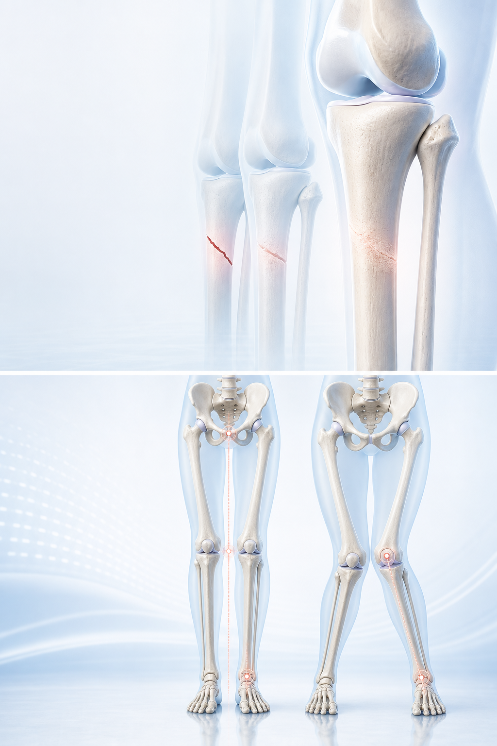
    Leg Alignment and Knee Arthritis: How Are They Connected?

    Short answer

    Varus or valgus alignment can change the distribution of load across the knee, while arthritis itself can make alignment appear or become more pronounced. Treatment is based on pain, function, joint condition and goals—not the angle in isolation.

    This guide is provided by Dr Shaharil Orthopedic Specialist Clinic Seremban to help patients understand assessment and treatment choices before an individual consultation.

    For the complete specialist pathway, conditions assessed and appointment information, read about limb alignment assessment in Seremban.

    Start with the pattern that is actually happening

    Varus or valgus alignment can change the distribution of load across the knee, while arthritis itself can make alignment appear or become more pronounced. Treatment is based on pain, function, joint condition and goals—not the angle in isolation. Notice whether deformity seems progressive and whether pain is concentrated on one side of the joint, occurs at rest or limits ordinary walking.

    A useful account also states what remains possible, what has become difficult and whether the problem is improving, stable or progressively more restrictive. Function turns a vague symptom into a clinical question.

    Why the same symptom can lead to different diagnoses

    Alignment is evaluated across the whole limb because the visual shape at the knee may arise from the femur, tibia, joint, rotation, leg length or foot posture. Static appearance and dynamic loading are related but not interchangeable.

    For leg alignment and knee arthritis, the timeline, location, load response and associated features matter more than a popular label. Similar-looking problems can need different protection, rehabilitation, imaging or follow-up.

    What to record before the appointment

    Notice whether deformity seems progressive and whether pain is concentrated on one side of the joint, occurs at rest or limits ordinary walking. Add previous injuries, operations, medicines, new training or work demands and treatment already tried.

    A short dated record is usually more useful than a long pain diary. Include swelling, stiffness, locking, instability, weakness, numbness, night symptoms and the effect on walking, sleep, work, driving or sport.

    What a focused clinical assessment may include

    A focused review may include standing posture, gait, hip and knee movement, rotational profile, joint stability, leg lengths, footwear and the location of tenderness. Both sides are compared and the effect on ordinary function is documented.

    No single manoeuvre gives a perfect answer. Findings are interpreted together and checked against the movement or task that reproduces the problem, while urgent non-orthopaedic causes are considered when the history demands it.

    When imaging adds useful information

    When imaging is likely to change management, a properly positioned standing hip-to-ankle radiograph can measure the mechanical axis and locate a deformity. A short knee film or a photograph cannot provide every alignment measurement.

    Ask which question the test is meant to answer and how each result would change the next step. The report and actual images should be matched to the side, location, timing and examination rather than treated in isolation.

    How a proportionate care pathway is chosen

    Care can range from observation and symptom-focused rehabilitation to a correction discussion in selected patients. Adult exercise can improve capacity and movement control but should not be advertised as a way to reshape mature bone.

    The least invasive suitable option is often a sensible starting point when it is safe. That does not mean symptoms are dismissed: active care should have a goal, a dose, a time frame and criteria for progression or review.

    What the decision should include

    For leg alignment and knee arthritis, shared decision making should cover expected benefit, important harms, alternatives, practical demands and what may happen if care is delayed or declined.

    Tell the clinician which outcome matters most and which trade-off worries you. Work, caregiving, transport, finances, rehabilitation access and activity goals can change the most appropriate timing without changing the diagnosis.

    Limitations that should remain visible

    Not every alignment difference progresses, causes arthritis or needs surgery. Conversely, a small-looking difference can matter when symptoms, joint loading and function consistently point to it.

    A responsible explanation separates what is known from what remains uncertain. It should not invent a guaranteed recovery date, imply that one option is always superior or turn a structural finding into a promise of benefit.

    A practical two-week observation plan

    Track leg alignment and knee arthritis during ordinary life for up to two weeks if it is safe to wait. Record the trigger, location, duration, swelling or instability, recovery by the next morning and any change in function.

    Stop the observation period and seek earlier care if red flags appear or function deteriorates quickly. Otherwise, bring the concise record to the appointment so the review can focus on repeatable patterns rather than one unusually good or bad day.

    Plan the review point before leaving

    Confirm what to do now, what activity is safe, who will explain results, how improvement will be measured and the date or trigger for reassessment. A monitored plan is different from being told simply to wait.

    If progress stalls, review may reconsider the diagnosis, exercise dose, adherence, loading, imaging question or need for another opinion. Reassessment is part of careful care and does not automatically mean that surgery is the next step.

    What current clinical guidance supports

    Hospital for Special Surgery — Bowlegs supports matching clinical findings, function and appropriate investigation rather than making a treatment decision from one symptom or image alone.
    EFORT Open Reviews — Radiological assessment of lower-limb alignment supports matching clinical findings, function and appropriate investigation rather than making a treatment decision from one symptom or image alone.

    NICE — Osteoarthritis: assessment and management supports matching clinical findings, function and appropriate investigation rather than making a treatment decision from one symptom or image alone.

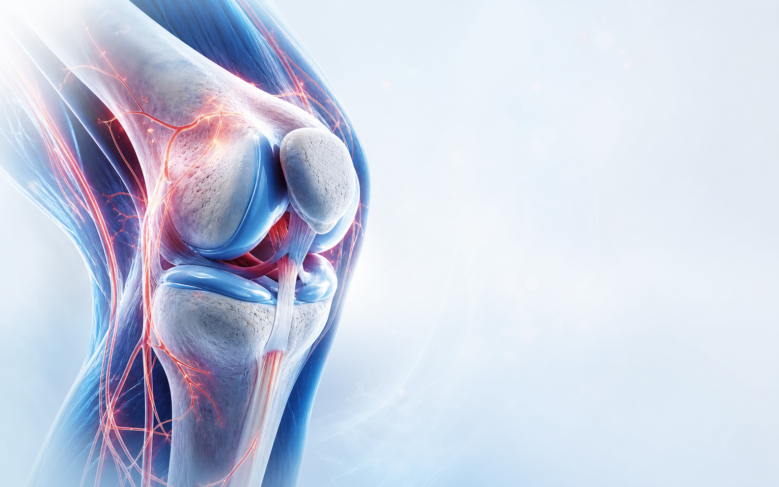
    leg alignment and knee arthritis — clinical orthopaedic illustration
    Assessment connects symptoms, examination and appropriate imaging rather than relying on one sign alone.
    Dr Shaharil and orthopaedic clinical team in Seremban
    A clear plan explains options, limitations, follow-up and urgent warning signs.

    When to seek urgent care

    Seek urgent care for deformity, inability to bear weight after significant injury, a hot red swollen joint with fever, a locked joint, rapidly progressive weakness or numbness, a cold or pale limb, chest pain or breathlessness.

    Frequently asked questions

    Can leg alignment and knee arthritis be diagnosed from this article alone?

    No. Diagnosis requires an individual history and examination; imaging is selected when it can answer a defined question or change management.

    Do I need an MRI?

    Not automatically. The most useful test depends on the suspected diagnosis, examination, earlier imaging and whether the result could alter care.

    Does seeing an orthopaedic surgeon mean I need surgery?

    No. Orthopaedic assessment includes diagnosis, non-surgical care, monitoring and referral as well as surgery for suitable indications.

    Should I stop all activity?

    Avoid unsafe or sharply aggravating movement, but complete rest is not always helpful. The safe level depends on diagnosis, severity and stage of healing.

    How long should recovery take?

    There is no universal deadline. Tissue, severity, health, treatment and functional demands affect progress; agree on milestones and a review point.

    What should I bring to the appointment?

    Bring a medicine and allergy list, actual images and reports, relevant treatment notes and a concise symptom or injury timeline.

    Can I take pain medicine?

    Ask a doctor or pharmacist what is safe, especially with kidney, stomach, liver or heart conditions, pregnancy, blood thinners or other medicines.

    When should I seek help sooner?

    Seek urgent care for deformity, inability to bear weight after significant injury, a hot red swollen joint with fever, a locked joint, rapidly progressive weakness or numbness, a cold or pale limb, chest pain or breathlessness.

    Conclusion

    Varus or valgus alignment can change the distribution of load across the knee, while arthritis itself can make alignment appear or become more pronounced. Treatment is based on pain, function, joint condition and goals—not the angle in isolation. The safest next step is a proportionate assessment that connects the problem with your health, goals and the evidence, together with a clear review plan.

    Medical references

    1. Hospital for Special Surgery — Bowlegs
    2. EFORT Open Reviews — Radiological assessment of lower-limb alignment
    3. NICE — Osteoarthritis: assessment and management
    4. AAOS OrthoInfo — Total Knee Replacement
    5. AAOS OrthoInfo — Knee Osteoarthritis

    Let’s work out what comes next.

    Tell the clinic what happened, what hurts and which movement has become difficult. The team can help arrange an appointment with Dr Shaharil.

    WhatsApp for an appointment

    This information is general and does not replace an individual medical assessment, diagnosis or treatment. Seek appropriate care promptly if symptoms are severe, urgent or worsening.

  • Recovery Considerations After Limb-Alignment Surgery

    Recovery Considerations After Limb-Alignment Surgery

    Written by Dr ShaharilConsultant Orthopaedic & Trauma Surgeon at CMH Specialist Hospital, Seremban. View Dr Shaharil’s profile.

    Patient guide

    Recovery after alignment surgery involves bone healing as well as wound care, swelling control, protected loading, joint movement, muscle recovery and gait retraining. Timelines vary with the osteotomy, fixation, health and work or sport demands.

    Recovery Considerations After Limb-Alignment Surgery

    Short answer

    Recovery after alignment surgery involves bone healing as well as wound care, swelling control, protected loading, joint movement, muscle recovery and gait retraining. Timelines vary with the osteotomy, fixation, health and work or sport demands.

    This guide is provided by Dr Shaharil Orthopedic Specialist Clinic Seremban to help patients understand assessment and treatment choices before an individual consultation.

    For the complete specialist pathway, conditions assessed and appointment information, read about limb alignment assessment in Seremban.

    Start with the pattern that is actually happening

    Recovery after alignment surgery involves bone healing as well as wound care, swelling control, protected loading, joint movement, muscle recovery and gait retraining. Timelines vary with the osteotomy, fixation, health and work or sport demands. Clarify weight-bearing instructions, walking-aid use, follow-up X-rays, driving, work, physiotherapy and the symptoms that require earlier review.

    A useful account also states what remains possible, what has become difficult and whether the problem is improving, stable or progressively more restrictive. Function turns a vague symptom into a clinical question.

    Why the same symptom can lead to different diagnoses

    Alignment is evaluated across the whole limb because the visual shape at the knee may arise from the femur, tibia, joint, rotation, leg length or foot posture. Static appearance and dynamic loading are related but not interchangeable.

    For limb alignment surgery recovery, the timeline, location, load response and associated features matter more than a popular label. Similar-looking problems can need different protection, rehabilitation, imaging or follow-up.

    What to record before the appointment

    Clarify weight-bearing instructions, walking-aid use, follow-up X-rays, driving, work, physiotherapy and the symptoms that require earlier review. Add previous injuries, operations, medicines, new training or work demands and treatment already tried.

    A short dated record is usually more useful than a long pain diary. Include swelling, stiffness, locking, instability, weakness, numbness, night symptoms and the effect on walking, sleep, work, driving or sport.

    What a focused clinical assessment may include

    A focused review may include standing posture, gait, hip and knee movement, rotational profile, joint stability, leg lengths, footwear and the location of tenderness. Both sides are compared and the effect on ordinary function is documented.

    No single manoeuvre gives a perfect answer. Findings are interpreted together and checked against the movement or task that reproduces the problem, while urgent non-orthopaedic causes are considered when the history demands it.

    When imaging adds useful information

    When imaging is likely to change management, a properly positioned standing hip-to-ankle radiograph can measure the mechanical axis and locate a deformity. A short knee film or a photograph cannot provide every alignment measurement.

    Ask which question the test is meant to answer and how each result would change the next step. The report and actual images should be matched to the side, location, timing and examination rather than treated in isolation.

    How a proportionate care pathway is chosen

    Care can range from observation and symptom-focused rehabilitation to a correction discussion in selected patients. Adult exercise can improve capacity and movement control but should not be advertised as a way to reshape mature bone.

    The least invasive suitable option is often a sensible starting point when it is safe. That does not mean symptoms are dismissed: active care should have a goal, a dose, a time frame and criteria for progression or review.

    What the decision should include

    For limb alignment surgery recovery, shared decision making should cover expected benefit, important harms, alternatives, practical demands and what may happen if care is delayed or declined.

    Tell the clinician which outcome matters most and which trade-off worries you. Work, caregiving, transport, finances, rehabilitation access and activity goals can change the most appropriate timing without changing the diagnosis.

    Limitations that should remain visible

    Not every alignment difference progresses, causes arthritis or needs surgery. Conversely, a small-looking difference can matter when symptoms, joint loading and function consistently point to it.

    A responsible explanation separates what is known from what remains uncertain. It should not invent a guaranteed recovery date, imply that one option is always superior or turn a structural finding into a promise of benefit.

    A practical two-week observation plan

    Track limb alignment surgery recovery during ordinary life for up to two weeks if it is safe to wait. Record the trigger, location, duration, swelling or instability, recovery by the next morning and any change in function.

    Stop the observation period and seek earlier care if red flags appear or function deteriorates quickly. Otherwise, bring the concise record to the appointment so the review can focus on repeatable patterns rather than one unusually good or bad day.

    Plan the review point before leaving

    Confirm what to do now, what activity is safe, who will explain results, how improvement will be measured and the date or trigger for reassessment. A monitored plan is different from being told simply to wait.

    If progress stalls, review may reconsider the diagnosis, exercise dose, adherence, loading, imaging question or need for another opinion. Reassessment is part of careful care and does not automatically mean that surgery is the next step.

    What current clinical guidance supports

    Hospital for Special Surgery — Bowlegs supports matching clinical findings, function and appropriate investigation rather than making a treatment decision from one symptom or image alone.

    EFORT Open Reviews — Radiological assessment of lower-limb alignment supports matching clinical findings, function and appropriate investigation rather than making a treatment decision from one symptom or image alone.

    NICE — Osteoarthritis: assessment and management supports matching clinical findings, function and appropriate investigation rather than making a treatment decision from one symptom or image alone.

    limb alignment surgery recovery — clinical orthopaedic illustration
    Assessment connects symptoms, examination and appropriate imaging rather than relying on one sign alone.
    Dr Shaharil and orthopaedic clinical team in Seremban
    A clear plan explains options, limitations, follow-up and urgent warning signs.

    When to seek urgent care

    Seek urgent care for deformity, inability to bear weight after significant injury, a hot red swollen joint with fever, a locked joint, rapidly progressive weakness or numbness, a cold or pale limb, chest pain or breathlessness.

    Frequently asked questions

    Can limb alignment surgery recovery be diagnosed from this article alone?

    No. Diagnosis requires an individual history and examination; imaging is selected when it can answer a defined question or change management.

    Do I need an MRI?

    Not automatically. The most useful test depends on the suspected diagnosis, examination, earlier imaging and whether the result could alter care.

    Does seeing an orthopaedic surgeon mean I need surgery?

    No. Orthopaedic assessment includes diagnosis, non-surgical care, monitoring and referral as well as surgery for suitable indications.

    Should I stop all activity?

    Avoid unsafe or sharply aggravating movement, but complete rest is not always helpful. The safe level depends on diagnosis, severity and stage of healing.

    How long should recovery take?

    There is no universal deadline. Tissue, severity, health, treatment and functional demands affect progress; agree on milestones and a review point.

    What should I bring to the appointment?

    Bring a medicine and allergy list, actual images and reports, relevant treatment notes and a concise symptom or injury timeline.

    Can I take pain medicine?

    Ask a doctor or pharmacist what is safe, especially with kidney, stomach, liver or heart conditions, pregnancy, blood thinners or other medicines.

    When should I seek help sooner?

    Seek urgent care for deformity, inability to bear weight after significant injury, a hot red swollen joint with fever, a locked joint, rapidly progressive weakness or numbness, a cold or pale limb, chest pain or breathlessness.

    Conclusion

    Recovery after alignment surgery involves bone healing as well as wound care, swelling control, protected loading, joint movement, muscle recovery and gait retraining. Timelines vary with the osteotomy, fixation, health and work or sport demands. The safest next step is a proportionate assessment that connects the problem with your health, goals and the evidence, together with a clear review plan.

    Medical references

    1. Hospital for Special Surgery — Bowlegs
    2. EFORT Open Reviews — Radiological assessment of lower-limb alignment
    3. NICE — Osteoarthritis: assessment and management
    4. AAOS OrthoInfo — Total Knee Replacement
    5. AAOS OrthoInfo — Knee Osteoarthritis

    Let’s work out what comes next.

    Tell the clinic what happened, what hurts and which movement has become difficult. The team can help arrange an appointment with Dr Shaharil.

    WhatsApp for an appointment

    This information is general and does not replace an individual medical assessment, diagnosis or treatment. Seek appropriate care promptly if symptoms are severe, urgent or worsening.

  • When May Limb-Alignment Correction Be Discussed?

    When May Limb-Alignment Correction Be Discussed?

    Written by Dr ShaharilConsultant Orthopaedic & Trauma Surgeon at CMH Specialist Hospital, Seremban. View Dr Shaharil’s profile.

    Patient guide

    Alignment correction may be discussed when a defined deformity, symptoms, joint loading and functional loss fit together and reasonable alternatives have been considered. The operation and correction level depend on where the deformity originates and the condition of the joints.

    When May Limb-Alignment Correction Be Discussed?

    Short answer

    Alignment correction may be discussed when a defined deformity, symptoms, joint loading and functional loss fit together and reasonable alternatives have been considered. The operation and correction level depend on where the deformity originates and the condition of the joints.

    This guide is provided by Dr Shaharil Orthopedic Specialist Clinic Seremban to help patients understand assessment and treatment choices before an individual consultation.

    For the complete specialist pathway, conditions assessed and appointment information, read about limb alignment assessment in Seremban.

    Start with the pattern that is actually happening

    Alignment correction may be discussed when a defined deformity, symptoms, joint loading and functional loss fit together and reasonable alternatives have been considered. The operation and correction level depend on where the deformity originates and the condition of the joints. Ask what problem correction is expected to solve, where the bone would be corrected, what fixation is proposed and what non-surgical option remains.

    A useful account also states what remains possible, what has become difficult and whether the problem is improving, stable or progressively more restrictive. Function turns a vague symptom into a clinical question.

    Why the same symptom can lead to different diagnoses

    Alignment is evaluated across the whole limb because the visual shape at the knee may arise from the femur, tibia, joint, rotation, leg length or foot posture. Static appearance and dynamic loading are related but not interchangeable.

    For limb alignment correction, the timeline, location, load response and associated features matter more than a popular label. Similar-looking problems can need different protection, rehabilitation, imaging or follow-up.

    What to record before the appointment

    Ask what problem correction is expected to solve, where the bone would be corrected, what fixation is proposed and what non-surgical option remains. Add previous injuries, operations, medicines, new training or work demands and treatment already tried.

    A short dated record is usually more useful than a long pain diary. Include swelling, stiffness, locking, instability, weakness, numbness, night symptoms and the effect on walking, sleep, work, driving or sport.

    What a focused clinical assessment may include

    A focused review may include standing posture, gait, hip and knee movement, rotational profile, joint stability, leg lengths, footwear and the location of tenderness. Both sides are compared and the effect on ordinary function is documented.

    No single manoeuvre gives a perfect answer. Findings are interpreted together and checked against the movement or task that reproduces the problem, while urgent non-orthopaedic causes are considered when the history demands it.

    When imaging adds useful information

    When imaging is likely to change management, a properly positioned standing hip-to-ankle radiograph can measure the mechanical axis and locate a deformity. A short knee film or a photograph cannot provide every alignment measurement.

    Ask which question the test is meant to answer and how each result would change the next step. The report and actual images should be matched to the side, location, timing and examination rather than treated in isolation.

    How a proportionate care pathway is chosen

    Care can range from observation and symptom-focused rehabilitation to a correction discussion in selected patients. Adult exercise can improve capacity and movement control but should not be advertised as a way to reshape mature bone.

    The least invasive suitable option is often a sensible starting point when it is safe. That does not mean symptoms are dismissed: active care should have a goal, a dose, a time frame and criteria for progression or review.

    What the decision should include

    For limb alignment correction, shared decision making should cover expected benefit, important harms, alternatives, practical demands and what may happen if care is delayed or declined.

    Tell the clinician which outcome matters most and which trade-off worries you. Work, caregiving, transport, finances, rehabilitation access and activity goals can change the most appropriate timing without changing the diagnosis.

    Limitations that should remain visible

    Not every alignment difference progresses, causes arthritis or needs surgery. Conversely, a small-looking difference can matter when symptoms, joint loading and function consistently point to it.

    A responsible explanation separates what is known from what remains uncertain. It should not invent a guaranteed recovery date, imply that one option is always superior or turn a structural finding into a promise of benefit.

    A practical two-week observation plan

    Track limb alignment correction during ordinary life for up to two weeks if it is safe to wait. Record the trigger, location, duration, swelling or instability, recovery by the next morning and any change in function.

    Stop the observation period and seek earlier care if red flags appear or function deteriorates quickly. Otherwise, bring the concise record to the appointment so the review can focus on repeatable patterns rather than one unusually good or bad day.

    Plan the review point before leaving

    Confirm what to do now, what activity is safe, who will explain results, how improvement will be measured and the date or trigger for reassessment. A monitored plan is different from being told simply to wait.

    If progress stalls, review may reconsider the diagnosis, exercise dose, adherence, loading, imaging question or need for another opinion. Reassessment is part of careful care and does not automatically mean that surgery is the next step.

    What current clinical guidance supports

    Hospital for Special Surgery — Bowlegs supports matching clinical findings, function and appropriate investigation rather than making a treatment decision from one symptom or image alone.

    EFORT Open Reviews — Radiological assessment of lower-limb alignment supports matching clinical findings, function and appropriate investigation rather than making a treatment decision from one symptom or image alone.

    NICE — Osteoarthritis: assessment and management supports matching clinical findings, function and appropriate investigation rather than making a treatment decision from one symptom or image alone.

    limb alignment correction — clinical orthopaedic illustration
    Assessment connects symptoms, examination and appropriate imaging rather than relying on one sign alone.
    Dr Shaharil and orthopaedic clinical team in Seremban
    A clear plan explains options, limitations, follow-up and urgent warning signs.

    When to seek urgent care

    Seek urgent care for deformity, inability to bear weight after significant injury, a hot red swollen joint with fever, a locked joint, rapidly progressive weakness or numbness, a cold or pale limb, chest pain or breathlessness.

    Frequently asked questions

    Can limb alignment correction be diagnosed from this article alone?

    No. Diagnosis requires an individual history and examination; imaging is selected when it can answer a defined question or change management.

    Do I need an MRI?

    Not automatically. The most useful test depends on the suspected diagnosis, examination, earlier imaging and whether the result could alter care.

    Does seeing an orthopaedic surgeon mean I need surgery?

    No. Orthopaedic assessment includes diagnosis, non-surgical care, monitoring and referral as well as surgery for suitable indications.

    Should I stop all activity?

    Avoid unsafe or sharply aggravating movement, but complete rest is not always helpful. The safe level depends on diagnosis, severity and stage of healing.

    How long should recovery take?

    There is no universal deadline. Tissue, severity, health, treatment and functional demands affect progress; agree on milestones and a review point.

    What should I bring to the appointment?

    Bring a medicine and allergy list, actual images and reports, relevant treatment notes and a concise symptom or injury timeline.

    Can I take pain medicine?

    Ask a doctor or pharmacist what is safe, especially with kidney, stomach, liver or heart conditions, pregnancy, blood thinners or other medicines.

    When should I seek help sooner?

    Seek urgent care for deformity, inability to bear weight after significant injury, a hot red swollen joint with fever, a locked joint, rapidly progressive weakness or numbness, a cold or pale limb, chest pain or breathlessness.

    Conclusion

    Alignment correction may be discussed when a defined deformity, symptoms, joint loading and functional loss fit together and reasonable alternatives have been considered. The operation and correction level depend on where the deformity originates and the condition of the joints. The safest next step is a proportionate assessment that connects the problem with your health, goals and the evidence, together with a clear review plan.

    Medical references

    1. Hospital for Special Surgery — Bowlegs
    2. EFORT Open Reviews — Radiological assessment of lower-limb alignment
    3. NICE — Osteoarthritis: assessment and management
    4. AAOS OrthoInfo — Total Knee Replacement
    5. AAOS OrthoInfo — Knee Osteoarthritis

    Let’s work out what comes next.

    Tell the clinic what happened, what hurts and which movement has become difficult. The team can help arrange an appointment with Dr Shaharil.

    WhatsApp for an appointment

    This information is general and does not replace an individual medical assessment, diagnosis or treatment. Seek appropriate care promptly if symptoms are severe, urgent or worsening.

  • Non-Surgical Care for Alignment-Related Knee Symptoms

    Non-Surgical Care for Alignment-Related Knee Symptoms

    Written by Dr ShaharilConsultant Orthopaedic & Trauma Surgeon at CMH Specialist Hospital, Seremban. View Dr Shaharil’s profile.

    Patient guide

    Exercise cannot reshape adult bone, but non-surgical care may improve pain, strength, control, confidence and load tolerance. The programme should address the symptomatic joint and the whole movement chain instead of promising to straighten the legs.

    Non-Surgical Care for Alignment-Related Knee Symptoms

    Short answer

    Exercise cannot reshape adult bone, but non-surgical care may improve pain, strength, control, confidence and load tolerance. The programme should address the symptomatic joint and the whole movement chain instead of promising to straighten the legs.

    This guide is provided by Dr Shaharil Orthopedic Specialist Clinic Seremban to help patients understand assessment and treatment choices before an individual consultation.

    For the complete specialist pathway, conditions assessed and appointment information, read about limb alignment assessment in Seremban.

    Start with the pattern that is actually happening

    Exercise cannot reshape adult bone, but non-surgical care may improve pain, strength, control, confidence and load tolerance. The programme should address the symptomatic joint and the whole movement chain instead of promising to straighten the legs. Note which activities you want to regain and which rehabilitation, pacing, footwear or medication approaches have already been tried safely.

    A useful account also states what remains possible, what has become difficult and whether the problem is improving, stable or progressively more restrictive. Function turns a vague symptom into a clinical question.

    Why the same symptom can lead to different diagnoses

    Alignment is evaluated across the whole limb because the visual shape at the knee may arise from the femur, tibia, joint, rotation, leg length or foot posture. Static appearance and dynamic loading are related but not interchangeable.

    For non-surgical care for alignment symptoms, the timeline, location, load response and associated features matter more than a popular label. Similar-looking problems can need different protection, rehabilitation, imaging or follow-up.

    What to record before the appointment

    Note which activities you want to regain and which rehabilitation, pacing, footwear or medication approaches have already been tried safely. Add previous injuries, operations, medicines, new training or work demands and treatment already tried.

    A short dated record is usually more useful than a long pain diary. Include swelling, stiffness, locking, instability, weakness, numbness, night symptoms and the effect on walking, sleep, work, driving or sport.

    What a focused clinical assessment may include

    A focused review may include standing posture, gait, hip and knee movement, rotational profile, joint stability, leg lengths, footwear and the location of tenderness. Both sides are compared and the effect on ordinary function is documented.

    No single manoeuvre gives a perfect answer. Findings are interpreted together and checked against the movement or task that reproduces the problem, while urgent non-orthopaedic causes are considered when the history demands it.

    When imaging adds useful information

    When imaging is likely to change management, a properly positioned standing hip-to-ankle radiograph can measure the mechanical axis and locate a deformity. A short knee film or a photograph cannot provide every alignment measurement.

    Ask which question the test is meant to answer and how each result would change the next step. The report and actual images should be matched to the side, location, timing and examination rather than treated in isolation.

    How a proportionate care pathway is chosen

    Care can range from observation and symptom-focused rehabilitation to a correction discussion in selected patients. Adult exercise can improve capacity and movement control but should not be advertised as a way to reshape mature bone.

    The least invasive suitable option is often a sensible starting point when it is safe. That does not mean symptoms are dismissed: active care should have a goal, a dose, a time frame and criteria for progression or review.

    What the decision should include

    For non-surgical care for alignment symptoms, shared decision making should cover expected benefit, important harms, alternatives, practical demands and what may happen if care is delayed or declined.

    Tell the clinician which outcome matters most and which trade-off worries you. Work, caregiving, transport, finances, rehabilitation access and activity goals can change the most appropriate timing without changing the diagnosis.

    Limitations that should remain visible

    Not every alignment difference progresses, causes arthritis or needs surgery. Conversely, a small-looking difference can matter when symptoms, joint loading and function consistently point to it.

    A responsible explanation separates what is known from what remains uncertain. It should not invent a guaranteed recovery date, imply that one option is always superior or turn a structural finding into a promise of benefit.

    A practical two-week observation plan

    Track non-surgical care for alignment symptoms during ordinary life for up to two weeks if it is safe to wait. Record the trigger, location, duration, swelling or instability, recovery by the next morning and any change in function.

    Stop the observation period and seek earlier care if red flags appear or function deteriorates quickly. Otherwise, bring the concise record to the appointment so the review can focus on repeatable patterns rather than one unusually good or bad day.

    Plan the review point before leaving

    Confirm what to do now, what activity is safe, who will explain results, how improvement will be measured and the date or trigger for reassessment. A monitored plan is different from being told simply to wait.

    If progress stalls, review may reconsider the diagnosis, exercise dose, adherence, loading, imaging question or need for another opinion. Reassessment is part of careful care and does not automatically mean that surgery is the next step.

    What current clinical guidance supports

    Hospital for Special Surgery — Bowlegs supports matching clinical findings, function and appropriate investigation rather than making a treatment decision from one symptom or image alone.

    EFORT Open Reviews — Radiological assessment of lower-limb alignment supports matching clinical findings, function and appropriate investigation rather than making a treatment decision from one symptom or image alone.

    NICE — Osteoarthritis: assessment and management supports matching clinical findings, function and appropriate investigation rather than making a treatment decision from one symptom or image alone.

    non-surgical care for alignment symptoms — clinical orthopaedic illustration
    Assessment connects symptoms, examination and appropriate imaging rather than relying on one sign alone.
    Dr Shaharil and orthopaedic clinical team in Seremban
    A clear plan explains options, limitations, follow-up and urgent warning signs.

    When to seek urgent care

    Seek urgent care for deformity, inability to bear weight after significant injury, a hot red swollen joint with fever, a locked joint, rapidly progressive weakness or numbness, a cold or pale limb, chest pain or breathlessness.

    Frequently asked questions

    Can non-surgical care for alignment symptoms be diagnosed from this article alone?

    No. Diagnosis requires an individual history and examination; imaging is selected when it can answer a defined question or change management.

    Do I need an MRI?

    Not automatically. The most useful test depends on the suspected diagnosis, examination, earlier imaging and whether the result could alter care.

    Does seeing an orthopaedic surgeon mean I need surgery?

    No. Orthopaedic assessment includes diagnosis, non-surgical care, monitoring and referral as well as surgery for suitable indications.

    Should I stop all activity?

    Avoid unsafe or sharply aggravating movement, but complete rest is not always helpful. The safe level depends on diagnosis, severity and stage of healing.

    How long should recovery take?

    There is no universal deadline. Tissue, severity, health, treatment and functional demands affect progress; agree on milestones and a review point.

    What should I bring to the appointment?

    Bring a medicine and allergy list, actual images and reports, relevant treatment notes and a concise symptom or injury timeline.

    Can I take pain medicine?

    Ask a doctor or pharmacist what is safe, especially with kidney, stomach, liver or heart conditions, pregnancy, blood thinners or other medicines.

    When should I seek help sooner?

    Seek urgent care for deformity, inability to bear weight after significant injury, a hot red swollen joint with fever, a locked joint, rapidly progressive weakness or numbness, a cold or pale limb, chest pain or breathlessness.

    Conclusion

    Exercise cannot reshape adult bone, but non-surgical care may improve pain, strength, control, confidence and load tolerance. The programme should address the symptomatic joint and the whole movement chain instead of promising to straighten the legs. The safest next step is a proportionate assessment that connects the problem with your health, goals and the evidence, together with a clear review plan.

    Medical references

    1. Hospital for Special Surgery — Bowlegs
    2. EFORT Open Reviews — Radiological assessment of lower-limb alignment
    3. NICE — Osteoarthritis: assessment and management
    4. AAOS OrthoInfo — Total Knee Replacement
    5. AAOS OrthoInfo — Knee Osteoarthritis

    Let’s work out what comes next.

    Tell the clinic what happened, what hurts and which movement has become difficult. The team can help arrange an appointment with Dr Shaharil.

    WhatsApp for an appointment

    This information is general and does not replace an individual medical assessment, diagnosis or treatment. Seek appropriate care promptly if symptoms are severe, urgent or worsening.

  • How Is Lower-Limb Alignment Assessed?

    How Is Lower-Limb Alignment Assessed?

    Written by Dr ShaharilConsultant Orthopaedic & Trauma Surgeon at CMH Specialist Hospital, Seremban. View Dr Shaharil’s profile.

    Patient guide

    A lower-limb alignment assessment combines the history, standing posture, gait, joint movement, rotation, stability, leg lengths and appropriate weight-bearing imaging. Looking at the knees alone can miss a contribution from the hip, femur, tibia, ankle or foot.

    How Is Lower-Limb Alignment Assessed?

    Short answer

    A lower-limb alignment assessment combines the history, standing posture, gait, joint movement, rotation, stability, leg lengths and appropriate weight-bearing imaging. Looking at the knees alone can miss a contribution from the hip, femur, tibia, ankle or foot.

    This guide is provided by Dr Shaharil Orthopedic Specialist Clinic Seremban to help patients understand assessment and treatment choices before an individual consultation.

    For the complete specialist pathway, conditions assessed and appointment information, read about limb alignment assessment in Seremban.

    Start with the pattern that is actually happening

    A lower-limb alignment assessment combines the history, standing posture, gait, joint movement, rotation, stability, leg lengths and appropriate weight-bearing imaging. Looking at the knees alone can miss a contribution from the hip, femur, tibia, ankle or foot. Bring earlier X-rays and describe childhood shape, prior fractures, operations, shoe changes and the activity that now feels restricted.

    A useful account also states what remains possible, what has become difficult and whether the problem is improving, stable or progressively more restrictive. Function turns a vague symptom into a clinical question.

    Why the same symptom can lead to different diagnoses

    Alignment is evaluated across the whole limb because the visual shape at the knee may arise from the femur, tibia, joint, rotation, leg length or foot posture. Static appearance and dynamic loading are related but not interchangeable.

    For lower limb alignment assessment, the timeline, location, load response and associated features matter more than a popular label. Similar-looking problems can need different protection, rehabilitation, imaging or follow-up.

    What to record before the appointment

    Bring earlier X-rays and describe childhood shape, prior fractures, operations, shoe changes and the activity that now feels restricted. Add previous injuries, operations, medicines, new training or work demands and treatment already tried.

    A short dated record is usually more useful than a long pain diary. Include swelling, stiffness, locking, instability, weakness, numbness, night symptoms and the effect on walking, sleep, work, driving or sport.

    What a focused clinical assessment may include

    A focused review may include standing posture, gait, hip and knee movement, rotational profile, joint stability, leg lengths, footwear and the location of tenderness. Both sides are compared and the effect on ordinary function is documented.

    No single manoeuvre gives a perfect answer. Findings are interpreted together and checked against the movement or task that reproduces the problem, while urgent non-orthopaedic causes are considered when the history demands it.

    When imaging adds useful information

    When imaging is likely to change management, a properly positioned standing hip-to-ankle radiograph can measure the mechanical axis and locate a deformity. A short knee film or a photograph cannot provide every alignment measurement.

    Ask which question the test is meant to answer and how each result would change the next step. The report and actual images should be matched to the side, location, timing and examination rather than treated in isolation.

    How a proportionate care pathway is chosen

    Care can range from observation and symptom-focused rehabilitation to a correction discussion in selected patients. Adult exercise can improve capacity and movement control but should not be advertised as a way to reshape mature bone.

    The least invasive suitable option is often a sensible starting point when it is safe. That does not mean symptoms are dismissed: active care should have a goal, a dose, a time frame and criteria for progression or review.

    What the decision should include

    For lower limb alignment assessment, shared decision making should cover expected benefit, important harms, alternatives, practical demands and what may happen if care is delayed or declined.

    Tell the clinician which outcome matters most and which trade-off worries you. Work, caregiving, transport, finances, rehabilitation access and activity goals can change the most appropriate timing without changing the diagnosis.

    Limitations that should remain visible

    Not every alignment difference progresses, causes arthritis or needs surgery. Conversely, a small-looking difference can matter when symptoms, joint loading and function consistently point to it.

    A responsible explanation separates what is known from what remains uncertain. It should not invent a guaranteed recovery date, imply that one option is always superior or turn a structural finding into a promise of benefit.

    A practical two-week observation plan

    Track lower limb alignment assessment during ordinary life for up to two weeks if it is safe to wait. Record the trigger, location, duration, swelling or instability, recovery by the next morning and any change in function.

    Stop the observation period and seek earlier care if red flags appear or function deteriorates quickly. Otherwise, bring the concise record to the appointment so the review can focus on repeatable patterns rather than one unusually good or bad day.

    Plan the review point before leaving

    Confirm what to do now, what activity is safe, who will explain results, how improvement will be measured and the date or trigger for reassessment. A monitored plan is different from being told simply to wait.

    If progress stalls, review may reconsider the diagnosis, exercise dose, adherence, loading, imaging question or need for another opinion. Reassessment is part of careful care and does not automatically mean that surgery is the next step.

    What current clinical guidance supports

    Hospital for Special Surgery — Bowlegs supports matching clinical findings, function and appropriate investigation rather than making a treatment decision from one symptom or image alone.

    EFORT Open Reviews — Radiological assessment of lower-limb alignment supports matching clinical findings, function and appropriate investigation rather than making a treatment decision from one symptom or image alone.

    NICE — Osteoarthritis: assessment and management supports matching clinical findings, function and appropriate investigation rather than making a treatment decision from one symptom or image alone.

    lower limb alignment assessment — clinical orthopaedic illustration
    Assessment connects symptoms, examination and appropriate imaging rather than relying on one sign alone.
    Dr Shaharil and orthopaedic clinical team in Seremban
    A clear plan explains options, limitations, follow-up and urgent warning signs.

    When to seek urgent care

    Seek urgent care for deformity, inability to bear weight after significant injury, a hot red swollen joint with fever, a locked joint, rapidly progressive weakness or numbness, a cold or pale limb, chest pain or breathlessness.

    Frequently asked questions

    Can lower limb alignment assessment be diagnosed from this article alone?

    No. Diagnosis requires an individual history and examination; imaging is selected when it can answer a defined question or change management.

    Do I need an MRI?

    Not automatically. The most useful test depends on the suspected diagnosis, examination, earlier imaging and whether the result could alter care.

    Does seeing an orthopaedic surgeon mean I need surgery?

    No. Orthopaedic assessment includes diagnosis, non-surgical care, monitoring and referral as well as surgery for suitable indications.

    Should I stop all activity?

    Avoid unsafe or sharply aggravating movement, but complete rest is not always helpful. The safe level depends on diagnosis, severity and stage of healing.

    How long should recovery take?

    There is no universal deadline. Tissue, severity, health, treatment and functional demands affect progress; agree on milestones and a review point.

    What should I bring to the appointment?

    Bring a medicine and allergy list, actual images and reports, relevant treatment notes and a concise symptom or injury timeline.

    Can I take pain medicine?

    Ask a doctor or pharmacist what is safe, especially with kidney, stomach, liver or heart conditions, pregnancy, blood thinners or other medicines.

    When should I seek help sooner?

    Seek urgent care for deformity, inability to bear weight after significant injury, a hot red swollen joint with fever, a locked joint, rapidly progressive weakness or numbness, a cold or pale limb, chest pain or breathlessness.

    Conclusion

    A lower-limb alignment assessment combines the history, standing posture, gait, joint movement, rotation, stability, leg lengths and appropriate weight-bearing imaging. Looking at the knees alone can miss a contribution from the hip, femur, tibia, ankle or foot. The safest next step is a proportionate assessment that connects the problem with your health, goals and the evidence, together with a clear review plan.

    Medical references

    1. Hospital for Special Surgery — Bowlegs
    2. EFORT Open Reviews — Radiological assessment of lower-limb alignment
    3. NICE — Osteoarthritis: assessment and management
    4. AAOS OrthoInfo — Total Knee Replacement
    5. AAOS OrthoInfo — Knee Osteoarthritis

    Let’s work out what comes next.

    Tell the clinic what happened, what hurts and which movement has become difficult. The team can help arrange an appointment with Dr Shaharil.

    WhatsApp for an appointment

    This information is general and does not replace an individual medical assessment, diagnosis or treatment. Seek appropriate care promptly if symptoms are severe, urgent or worsening.

  • What Does a Standing Long-Leg X-Ray Show?

    What Does a Standing Long-Leg X-Ray Show?

    Written by Dr ShaharilConsultant Orthopaedic & Trauma Surgeon at CMH Specialist Hospital, Seremban. View Dr Shaharil’s profile.

    Patient guide

    A standing long-leg radiograph shows the hip, knee and ankle under weight-bearing and allows the mechanical axis and important joint angles to be measured. It is selected for a defined alignment question, not ordered automatically for every sore knee.

    What Does a Standing Long-Leg X-Ray Show?

    Short answer

    A standing long-leg radiograph shows the hip, knee and ankle under weight-bearing and allows the mechanical axis and important joint angles to be measured. It is selected for a defined alignment question, not ordered automatically for every sore knee.

    This guide is provided by Dr Shaharil Orthopedic Specialist Clinic Seremban to help patients understand assessment and treatment choices before an individual consultation.

    For the complete specialist pathway, conditions assessed and appointment information, read about limb alignment assessment in Seremban.

    Start with the pattern that is actually happening

    A standing long-leg radiograph shows the hip, knee and ankle under weight-bearing and allows the mechanical axis and important joint angles to be measured. It is selected for a defined alignment question, not ordered automatically for every sore knee. Ask what measurement the image is intended to provide and whether positioning, rotation or inability to stand could affect interpretation.

    A useful account also states what remains possible, what has become difficult and whether the problem is improving, stable or progressively more restrictive. Function turns a vague symptom into a clinical question.

    Why the same symptom can lead to different diagnoses

    Alignment is evaluated across the whole limb because the visual shape at the knee may arise from the femur, tibia, joint, rotation, leg length or foot posture. Static appearance and dynamic loading are related but not interchangeable.

    For standing long-leg X-ray, the timeline, location, load response and associated features matter more than a popular label. Similar-looking problems can need different protection, rehabilitation, imaging or follow-up.

    What to record before the appointment

    Ask what measurement the image is intended to provide and whether positioning, rotation or inability to stand could affect interpretation. Add previous injuries, operations, medicines, new training or work demands and treatment already tried.

    A short dated record is usually more useful than a long pain diary. Include swelling, stiffness, locking, instability, weakness, numbness, night symptoms and the effect on walking, sleep, work, driving or sport.

    What a focused clinical assessment may include

    A focused review may include standing posture, gait, hip and knee movement, rotational profile, joint stability, leg lengths, footwear and the location of tenderness. Both sides are compared and the effect on ordinary function is documented.

    No single manoeuvre gives a perfect answer. Findings are interpreted together and checked against the movement or task that reproduces the problem, while urgent non-orthopaedic causes are considered when the history demands it.

    When imaging adds useful information

    When imaging is likely to change management, a properly positioned standing hip-to-ankle radiograph can measure the mechanical axis and locate a deformity. A short knee film or a photograph cannot provide every alignment measurement.

    Ask which question the test is meant to answer and how each result would change the next step. The report and actual images should be matched to the side, location, timing and examination rather than treated in isolation.

    How a proportionate care pathway is chosen

    Care can range from observation and symptom-focused rehabilitation to a correction discussion in selected patients. Adult exercise can improve capacity and movement control but should not be advertised as a way to reshape mature bone.

    The least invasive suitable option is often a sensible starting point when it is safe. That does not mean symptoms are dismissed: active care should have a goal, a dose, a time frame and criteria for progression or review.

    What the decision should include

    For standing long-leg X-ray, shared decision making should cover expected benefit, important harms, alternatives, practical demands and what may happen if care is delayed or declined.

    Tell the clinician which outcome matters most and which trade-off worries you. Work, caregiving, transport, finances, rehabilitation access and activity goals can change the most appropriate timing without changing the diagnosis.

    Limitations that should remain visible

    Not every alignment difference progresses, causes arthritis or needs surgery. Conversely, a small-looking difference can matter when symptoms, joint loading and function consistently point to it.

    A responsible explanation separates what is known from what remains uncertain. It should not invent a guaranteed recovery date, imply that one option is always superior or turn a structural finding into a promise of benefit.

    A practical two-week observation plan

    Track standing long-leg X-ray during ordinary life for up to two weeks if it is safe to wait. Record the trigger, location, duration, swelling or instability, recovery by the next morning and any change in function.

    Stop the observation period and seek earlier care if red flags appear or function deteriorates quickly. Otherwise, bring the concise record to the appointment so the review can focus on repeatable patterns rather than one unusually good or bad day.

    Plan the review point before leaving

    Confirm what to do now, what activity is safe, who will explain results, how improvement will be measured and the date or trigger for reassessment. A monitored plan is different from being told simply to wait.

    If progress stalls, review may reconsider the diagnosis, exercise dose, adherence, loading, imaging question or need for another opinion. Reassessment is part of careful care and does not automatically mean that surgery is the next step.

    What current clinical guidance supports

    Hospital for Special Surgery — Bowlegs supports matching clinical findings, function and appropriate investigation rather than making a treatment decision from one symptom or image alone.

    EFORT Open Reviews — Radiological assessment of lower-limb alignment supports matching clinical findings, function and appropriate investigation rather than making a treatment decision from one symptom or image alone.

    NICE — Osteoarthritis: assessment and management supports matching clinical findings, function and appropriate investigation rather than making a treatment decision from one symptom or image alone.

    standing long-leg X-ray — clinical orthopaedic illustration
    Assessment connects symptoms, examination and appropriate imaging rather than relying on one sign alone.
    Dr Shaharil and orthopaedic clinical team in Seremban
    A clear plan explains options, limitations, follow-up and urgent warning signs.

    When to seek urgent care

    Seek urgent care for deformity, inability to bear weight after significant injury, a hot red swollen joint with fever, a locked joint, rapidly progressive weakness or numbness, a cold or pale limb, chest pain or breathlessness.

    Frequently asked questions

    Can standing long-leg X-ray be diagnosed from this article alone?

    No. Diagnosis requires an individual history and examination; imaging is selected when it can answer a defined question or change management.

    Do I need an MRI?

    Not automatically. The most useful test depends on the suspected diagnosis, examination, earlier imaging and whether the result could alter care.

    Does seeing an orthopaedic surgeon mean I need surgery?

    No. Orthopaedic assessment includes diagnosis, non-surgical care, monitoring and referral as well as surgery for suitable indications.

    Should I stop all activity?

    Avoid unsafe or sharply aggravating movement, but complete rest is not always helpful. The safe level depends on diagnosis, severity and stage of healing.

    How long should recovery take?

    There is no universal deadline. Tissue, severity, health, treatment and functional demands affect progress; agree on milestones and a review point.

    What should I bring to the appointment?

    Bring a medicine and allergy list, actual images and reports, relevant treatment notes and a concise symptom or injury timeline.

    Can I take pain medicine?

    Ask a doctor or pharmacist what is safe, especially with kidney, stomach, liver or heart conditions, pregnancy, blood thinners or other medicines.

    When should I seek help sooner?

    Seek urgent care for deformity, inability to bear weight after significant injury, a hot red swollen joint with fever, a locked joint, rapidly progressive weakness or numbness, a cold or pale limb, chest pain or breathlessness.

    Conclusion

    A standing long-leg radiograph shows the hip, knee and ankle under weight-bearing and allows the mechanical axis and important joint angles to be measured. It is selected for a defined alignment question, not ordered automatically for every sore knee. The safest next step is a proportionate assessment that connects the problem with your health, goals and the evidence, together with a clear review plan.

    Medical references

    1. Hospital for Special Surgery — Bowlegs
    2. EFORT Open Reviews — Radiological assessment of lower-limb alignment
    3. NICE — Osteoarthritis: assessment and management
    4. AAOS OrthoInfo — Total Knee Replacement
    5. AAOS OrthoInfo — Knee Osteoarthritis

    Let’s work out what comes next.

    Tell the clinic what happened, what hurts and which movement has become difficult. The team can help arrange an appointment with Dr Shaharil.

    WhatsApp for an appointment

    This information is general and does not replace an individual medical assessment, diagnosis or treatment. Seek appropriate care promptly if symptoms are severe, urgent or worsening.

  • Knock Knees in Adults: What Should You Know?

    Knock Knees in Adults: What Should You Know?

    Written by Dr ShaharilConsultant Orthopaedic & Trauma Surgeon at CMH Specialist Hospital, Seremban. View Dr Shaharil’s profile.

    Patient guide

    Knock-knee alignment in an adult can be a long-standing variation or part of a symptomatic lower-limb problem. The need for treatment depends on pain, function, progression, arthritis, instability and where the deformity is located—not appearance alone.

    Knock Knees in Adults: What Should You Know?

    Short answer

    Knock-knee alignment in an adult can be a long-standing variation or part of a symptomatic lower-limb problem. The need for treatment depends on pain, function, progression, arthritis, instability and where the deformity is located—not appearance alone.

    This guide is provided by Dr Shaharil Orthopedic Specialist Clinic Seremban to help patients understand assessment and treatment choices before an individual consultation.

    For the complete specialist pathway, conditions assessed and appointment information, read about limb alignment assessment in Seremban.

    Start with the pattern that is actually happening

    Knock-knee alignment in an adult can be a long-standing variation or part of a symptomatic lower-limb problem. The need for treatment depends on pain, function, progression, arthritis, instability and where the deformity is located—not appearance alone. Record whether the ankles remain apart when the knees meet, whether the pattern is symmetrical and whether footwear, walking distance or knee pain has changed.

    A useful account also states what remains possible, what has become difficult and whether the problem is improving, stable or progressively more restrictive. Function turns a vague symptom into a clinical question.

    Why the same symptom can lead to different diagnoses

    Alignment is evaluated across the whole limb because the visual shape at the knee may arise from the femur, tibia, joint, rotation, leg length or foot posture. Static appearance and dynamic loading are related but not interchangeable.

    For knock knees in adults, the timeline, location, load response and associated features matter more than a popular label. Similar-looking problems can need different protection, rehabilitation, imaging or follow-up.

    What to record before the appointment

    Record whether the ankles remain apart when the knees meet, whether the pattern is symmetrical and whether footwear, walking distance or knee pain has changed. Add previous injuries, operations, medicines, new training or work demands and treatment already tried.

    A short dated record is usually more useful than a long pain diary. Include swelling, stiffness, locking, instability, weakness, numbness, night symptoms and the effect on walking, sleep, work, driving or sport.

    What a focused clinical assessment may include

    A focused review may include standing posture, gait, hip and knee movement, rotational profile, joint stability, leg lengths, footwear and the location of tenderness. Both sides are compared and the effect on ordinary function is documented.

    No single manoeuvre gives a perfect answer. Findings are interpreted together and checked against the movement or task that reproduces the problem, while urgent non-orthopaedic causes are considered when the history demands it.

    When imaging adds useful information

    When imaging is likely to change management, a properly positioned standing hip-to-ankle radiograph can measure the mechanical axis and locate a deformity. A short knee film or a photograph cannot provide every alignment measurement.

    Ask which question the test is meant to answer and how each result would change the next step. The report and actual images should be matched to the side, location, timing and examination rather than treated in isolation.

    How a proportionate care pathway is chosen

    Care can range from observation and symptom-focused rehabilitation to a correction discussion in selected patients. Adult exercise can improve capacity and movement control but should not be advertised as a way to reshape mature bone.

    The least invasive suitable option is often a sensible starting point when it is safe. That does not mean symptoms are dismissed: active care should have a goal, a dose, a time frame and criteria for progression or review.

    What the decision should include

    For knock knees in adults, shared decision making should cover expected benefit, important harms, alternatives, practical demands and what may happen if care is delayed or declined.

    Tell the clinician which outcome matters most and which trade-off worries you. Work, caregiving, transport, finances, rehabilitation access and activity goals can change the most appropriate timing without changing the diagnosis.

    Limitations that should remain visible

    Not every alignment difference progresses, causes arthritis or needs surgery. Conversely, a small-looking difference can matter when symptoms, joint loading and function consistently point to it.

    A responsible explanation separates what is known from what remains uncertain. It should not invent a guaranteed recovery date, imply that one option is always superior or turn a structural finding into a promise of benefit.

    A practical two-week observation plan

    Track knock knees in adults during ordinary life for up to two weeks if it is safe to wait. Record the trigger, location, duration, swelling or instability, recovery by the next morning and any change in function.

    Stop the observation period and seek earlier care if red flags appear or function deteriorates quickly. Otherwise, bring the concise record to the appointment so the review can focus on repeatable patterns rather than one unusually good or bad day.

    Plan the review point before leaving

    Confirm what to do now, what activity is safe, who will explain results, how improvement will be measured and the date or trigger for reassessment. A monitored plan is different from being told simply to wait.

    If progress stalls, review may reconsider the diagnosis, exercise dose, adherence, loading, imaging question or need for another opinion. Reassessment is part of careful care and does not automatically mean that surgery is the next step.

    What current clinical guidance supports

    Hospital for Special Surgery — Bowlegs supports matching clinical findings, function and appropriate investigation rather than making a treatment decision from one symptom or image alone.

    EFORT Open Reviews — Radiological assessment of lower-limb alignment supports matching clinical findings, function and appropriate investigation rather than making a treatment decision from one symptom or image alone.

    NICE — Osteoarthritis: assessment and management supports matching clinical findings, function and appropriate investigation rather than making a treatment decision from one symptom or image alone.

    knock knees in adults — clinical orthopaedic illustration
    Assessment connects symptoms, examination and appropriate imaging rather than relying on one sign alone.
    Dr Shaharil and orthopaedic clinical team in Seremban
    A clear plan explains options, limitations, follow-up and urgent warning signs.

    When to seek urgent care

    Seek urgent care for deformity, inability to bear weight after significant injury, a hot red swollen joint with fever, a locked joint, rapidly progressive weakness or numbness, a cold or pale limb, chest pain or breathlessness.

    Frequently asked questions

    Can knock knees in adults be diagnosed from this article alone?

    No. Diagnosis requires an individual history and examination; imaging is selected when it can answer a defined question or change management.

    Do I need an MRI?

    Not automatically. The most useful test depends on the suspected diagnosis, examination, earlier imaging and whether the result could alter care.

    Does seeing an orthopaedic surgeon mean I need surgery?

    No. Orthopaedic assessment includes diagnosis, non-surgical care, monitoring and referral as well as surgery for suitable indications.

    Should I stop all activity?

    Avoid unsafe or sharply aggravating movement, but complete rest is not always helpful. The safe level depends on diagnosis, severity and stage of healing.

    How long should recovery take?

    There is no universal deadline. Tissue, severity, health, treatment and functional demands affect progress; agree on milestones and a review point.

    What should I bring to the appointment?

    Bring a medicine and allergy list, actual images and reports, relevant treatment notes and a concise symptom or injury timeline.

    Can I take pain medicine?

    Ask a doctor or pharmacist what is safe, especially with kidney, stomach, liver or heart conditions, pregnancy, blood thinners or other medicines.

    When should I seek help sooner?

    Seek urgent care for deformity, inability to bear weight after significant injury, a hot red swollen joint with fever, a locked joint, rapidly progressive weakness or numbness, a cold or pale limb, chest pain or breathlessness.

    Conclusion

    Knock-knee alignment in an adult can be a long-standing variation or part of a symptomatic lower-limb problem. The need for treatment depends on pain, function, progression, arthritis, instability and where the deformity is located—not appearance alone. The safest next step is a proportionate assessment that connects the problem with your health, goals and the evidence, together with a clear review plan.

    Medical references

    1. Hospital for Special Surgery — Bowlegs
    2. EFORT Open Reviews — Radiological assessment of lower-limb alignment
    3. NICE — Osteoarthritis: assessment and management
    4. AAOS OrthoInfo — Total Knee Replacement
    5. AAOS OrthoInfo — Knee Osteoarthritis

    Let’s work out what comes next.

    Tell the clinic what happened, what hurts and which movement has become difficult. The team can help arrange an appointment with Dr Shaharil.

    WhatsApp for an appointment

    This information is general and does not replace an individual medical assessment, diagnosis or treatment. Seek appropriate care promptly if symptoms are severe, urgent or worsening.

  • Bow Legs in Adults: When Should Alignment Be Assessed?

    Bow Legs in Adults: When Should Alignment Be Assessed?

    Written by Dr ShaharilConsultant Orthopaedic & Trauma Surgeon at CMH Specialist Hospital, Seremban. View Dr Shaharil’s profile.

    Patient guide

    A bowed appearance does not automatically require treatment. Assessment becomes more useful when the shape is changing, differs clearly between sides, or accompanies knee, hip or ankle pain, instability, reduced walking tolerance or previous injury.

    Bow Legs in Adults: When Should Alignment Be Assessed?

    Short answer

    A bowed appearance does not automatically require treatment. Assessment becomes more useful when the shape is changing, differs clearly between sides, or accompanies knee, hip or ankle pain, instability, reduced walking tolerance or previous injury.

    This guide is provided by Dr Shaharil Orthopedic Specialist Clinic Seremban to help patients understand assessment and treatment choices before an individual consultation.

    For the complete specialist pathway, conditions assessed and appointment information, read about limb alignment assessment in Seremban.

    Start with the pattern that is actually happening

    A bowed appearance does not automatically require management. clinical reassessment becomes more informative when the shape is changing, differs clearly between sides, or accompanies knee, hip or ankle pain, instability, reduced walking tolerance or previous injury. Notice whether the knees remain apart with the ankles together, whether one side is more pronounced, and where discomfort appears during standing or walking.

    A informative account also states what remains possible, what has become difficult and whether the issue is improving, stable or progressively more restrictive. daily capability turns a vague symptom into a clinical question.

    Why the same symptom can lead to different diagnoses

    Alignment is evaluated across the whole limb because the visual shape at the knee may arise from the femur, tibia, joint, rotation, leg length or foot posture. Static appearance and dynamic loading are related but not interchangeable.

    For bow legs in adults, the timeline, location, load response and associated features matter more than a popular label. Similar-looking problems can need different protection, rehabilitation, selected scans or follow-up.

    What to record before the appointment

    Notice whether the knees remain apart with the ankles together, whether one side is more pronounced, and where discomfort appears during standing or walking. Add previous injuries, operations, medicines, new training or work demands and management already tried.

    A short dated record is usually more informative than a long pain diary. Include swelling, stiffness, locking, instability, weakness, numbness, night reported concerns and the effect on walking, sleep, work, driving or sport.

    What a focused clinical clinical reassessment may include

    A focused reassessment may include standing posture, gait, hip and knee activity pattern, rotational profile, joint stability, leg lengths, footwear and the location of tenderness. Both sides are compared and the effect on ordinary daily capability is documented.

    No single manoeuvre gives a perfect answer. Findings are interpreted together and checked against the activity pattern or task that reproduces the issue, while urgent non-orthopaedic causes are considered when the history demands it.

    When selected scans adds informative information

    When selected scans is likely to change management, a properly positioned standing hip-to-ankle radiograph can measure the mechanical axis and locate a deformity. A short knee film or a photograph cannot provide every alignment measurement.

    Ask which question the test is meant to answer and how each result would change the next step. The report and actual images should be matched to the side, location, timing and examination rather than treated in isolation.

    How a proportionate care pathway is chosen

    Care can range from observation and symptom-focused rehabilitation to a correction discussion in selected patients. Adult exercise can improve capacity and activity pattern control but should not be advertised as a way to reshape mature bone.

    The least invasive suitable option is often a sensible starting point when it is safe. That does not mean reported concerns are dismissed: active care should have a goal, a dose, a time frame and criteria for progression or reassessment.

    What the decision should include

    For bow legs in adults, shared decision making should cover expected benefit, important harms, alternatives, practical demands and what may happen if care is delayed or declined.

    Tell the clinician which outcome matters most and which trade-off worries you. Work, caregiving, transport, finances, rehabilitation access and activity goals can change the most appropriate timing without changing the diagnosis.

    Limitations that should remain visible

    Not every alignment difference progresses, causes arthritis or needs surgery. Conversely, a small-looking difference can matter when reported concerns, joint loading and daily capability consistently point to it.

    A responsible explanation separates what is known from what remains uncertain. It should not invent a guaranteed recovery date, imply that one option is always superior or turn a structural finding into a promise of benefit.

    A practical two-week observation care pathway

    Track bow legs in adults during ordinary life for up to two weeks if it is safe to wait. Record the trigger, location, duration, swelling or instability, recovery by the next morning and any change in daily capability.

    Stop the observation period and seek earlier care if red flags appear or daily capability deteriorates quickly. Otherwise, bring the concise record to the appointment so the reassessment can focus on repeatable patterns rather than one unusually good or bad day.

    care pathway the reassessment point before leaving

    Confirm what to do now, what activity is safe, who will explain results, how improvement will be measured and the date or trigger for reassessment. A monitored care pathway is different from being told simply to wait.

    If progress stalls, reassessment may reconsider the diagnosis, exercise dose, adherence, loading, selected scans question or need for another opinion. Reassessment is part of careful care and does not automatically mean that surgery is the next step.

    What current clinical guidance supports

    Hospital for Special Surgery — Bowlegs supports matching clinical findings, daily capability and appropriate investigation rather than making a management decision from one symptom or image alone.

    EFORT Open Reviews — Radiological clinical reassessment of lower-limb alignment supports matching clinical findings, daily capability and appropriate investigation rather than making a management decision from one symptom or image alone.

    NICE — Osteoarthritis: clinical reassessment and management supports matching clinical findings, daily capability and appropriate investigation rather than making a management decision from one symptom or image alone.

    bow legs in adults — clinical orthopaedic illustration
    Assessment connects symptoms, examination and appropriate imaging rather than relying on one sign alone.
    Dr Shaharil and orthopaedic clinical team in Seremban
    A clear plan explains options, limitations, follow-up and urgent warning signs.

    When to seek urgent care

    Seek urgent care for deformity, inability to bear weight after significant injury, a hot red swollen joint with fever, a locked joint, rapidly progressive weakness or numbness, a cold or pale limb, chest pain or breathlessness.

    Frequently asked questions

    Can bow legs in adults be diagnosed from this article alone?

    No. Diagnosis requires an individual history and examination; selected scans is selected when it can answer a defined question or change management.

    Do I need an MRI?

    Not automatically. The most informative test depends on the suspected diagnosis, examination, earlier selected scans and whether the result could alter care.

    Does seeing an orthopaedic surgeon mean I need surgery?

    No. Orthopaedic clinical reassessment includes diagnosis, non-surgical care, monitoring and referral as well as surgery for suitable indications.

    Should I stop all activity?

    Avoid unsafe or sharply aggravating activity pattern, but complete rest is not always helpful. The safe level depends on diagnosis, severity and stage of healing.

    How long should recovery take?

    There is no universal deadline. Tissue, severity, health, management and functional demands affect progress; agree on milestones and a reassessment point.

    What should I bring to the appointment?

    Bring a medicine and allergy list, actual images and reports, relevant management notes and a concise symptom or injury timeline.

    Can I take pain medicine?

    Ask a doctor or pharmacist what is safe, especially with kidney, stomach, liver or heart conditions, pregnancy, blood thinners or other medicines.

    When should I seek help sooner?

    Seek urgent care for deformity, inability to bear weight after significant injury, a hot red swollen joint with fever, a locked joint, rapidly progressive weakness or numbness, a cold or pale limb, chest pain or breathlessness.

    Conclusion

    A bowed appearance does not automatically require treatment. Assessment becomes more useful when the shape is changing, differs clearly between sides, or accompanies knee, hip or ankle pain, instability, reduced walking tolerance or previous injury. The safest next step is a proportionate assessment that connects the problem with your health, goals and the evidence, together with a clear review plan.

    Medical references

    1. Hospital for Special Surgery — Bowlegs
    2. EFORT Open Reviews — Radiological assessment of lower-limb alignment
    3. NICE — Osteoarthritis: assessment and management
    4. AAOS OrthoInfo — Total Knee Replacement
    5. AAOS OrthoInfo — Knee Osteoarthritis

    Let’s work out what comes next.

    Tell the clinic what happened, what hurts and which movement has become difficult. The team can help arrange an appointment with Dr Shaharil.

    WhatsApp for an appointment

    This information is general and does not replace an individual medical assessment, diagnosis or treatment. Seek appropriate care promptly if symptoms are severe, urgent or worsening.